SKU: 16-0404
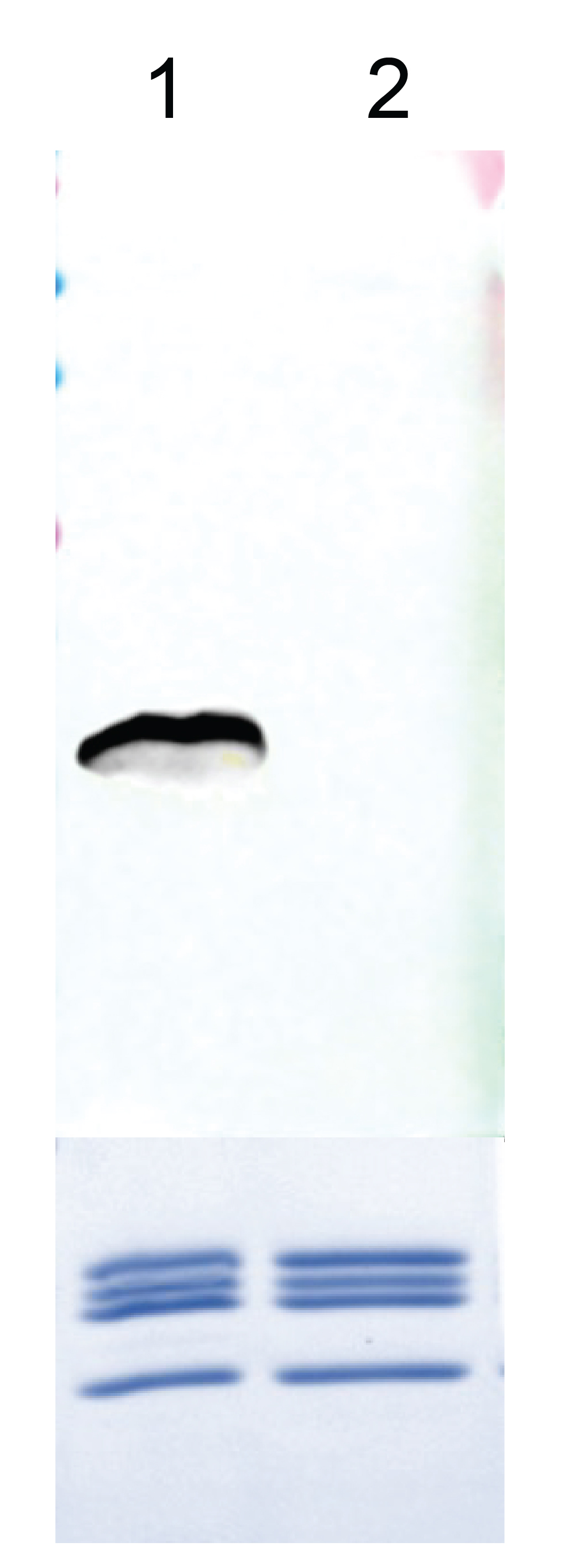
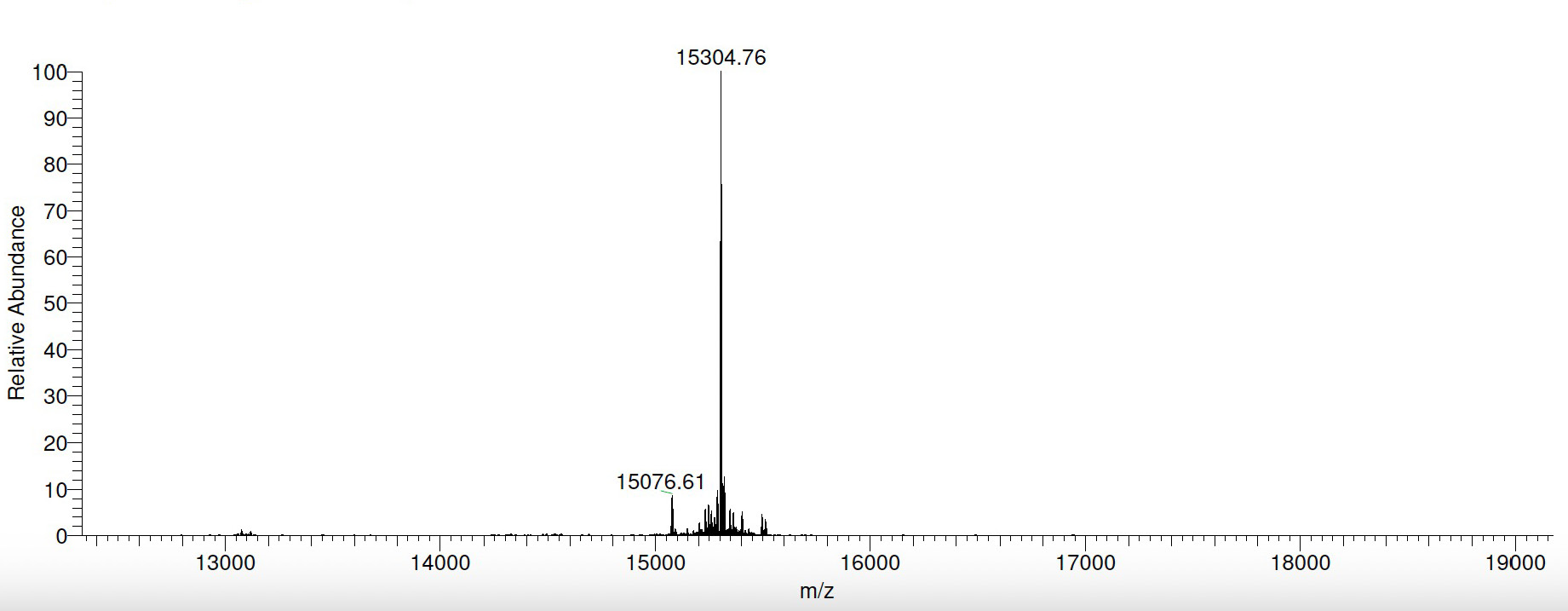
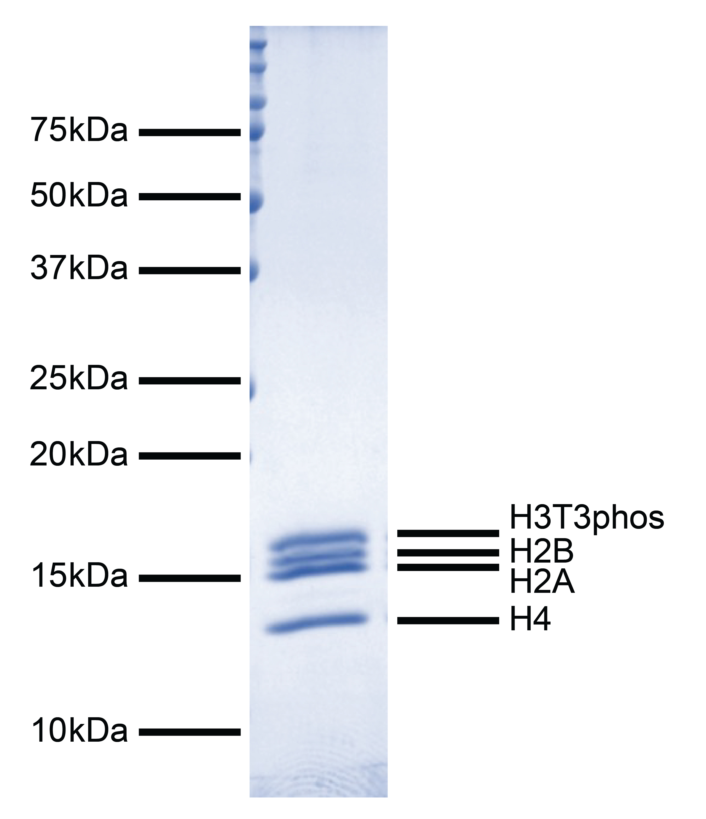
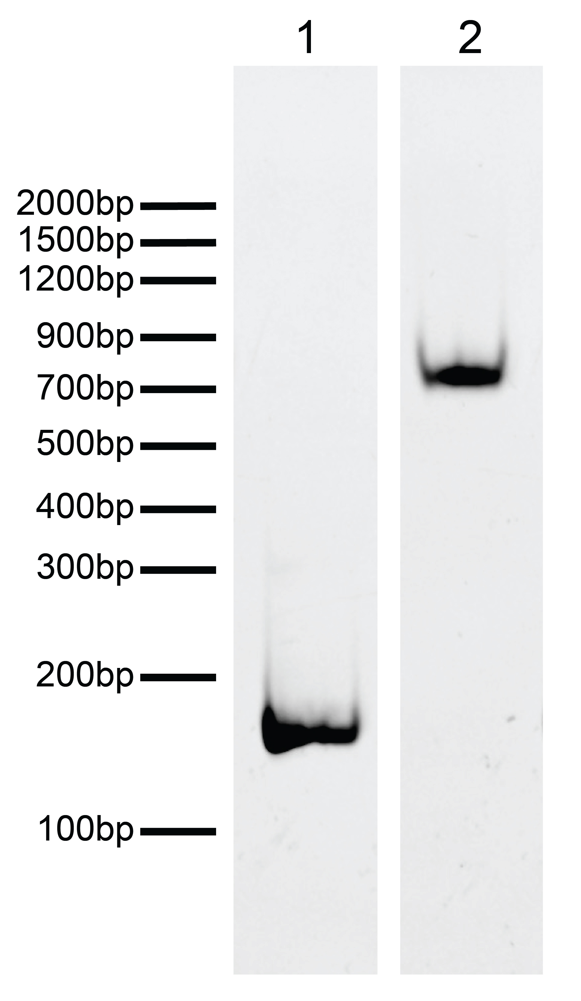
{"url":"https://www.epicypher.com/products/nucleosomes/modified-designer-nucleosomes-dnucs/h3t3phos-recombinant-nucleosome-biotinylated","add_this":[{"service":"facebook","annotation":""},{"service":"email","annotation":""},{"service":"print","annotation":""},{"service":"twitter","annotation":""},{"service":"linkedin","annotation":""}],"gtin":null,"options":[],"id":1149,"bulk_discount_rates":[],"can_purchase":true,"meta_description":"Phosphorylated designer nucleosome H3T3phos is highly purified and suitable for a variety of applications, including use as a substrate in enzyme assays, high-throughput screening and inhibitor testing, chromatin binding studies, protein-protein interaction assays, structural studies, and in effector protein binding experiments.","category":["Nucleosomes","Nucleosomes/Modified Designer Nucleosomes (dNucs™)"],"AddThisServiceButtonMeta":"","main_image":{"data":"https://cdn11.bigcommerce.com/s-y9o92/images/stencil/{:size}/products/1149/1223/Recombinant_nucleosome__92972.1717610733.png?c=2","alt":"H3T3phos Recombinant Nucleosome, Biotinylated"},"add_to_wishlist_url":"/wishlist.php?action=add&product_id=1149","shipping":{"calculated":true},"num_reviews":0,"weight":"0.01 LBS","custom_fields":[{"id":"1316","name":"Pack Size","value":"50 μg"}],"sku":"16-0404","description":"<div class=\"product-general-info\">\n <ul class=\"product-general-info__list-left\">\n <li class=\"product-general-info__list-item\">\n <strong>Species: </strong>Human\n </li>\n <li class=\"product-general-info__list-item\">\n <strong>Source: </strong><i>E. coli</i> & synthetic DNA\n </li>\n </ul>\n <ul class=\"product-general-info__list-right\">\n <li class=\"product-general-info__list-item\">\n <strong>Tag: </strong>Biotinylated\n </li>\n <li class=\"product-general-info__list-item\">\n <strong>Molecular Weight: </strong>199,806 Da\n </li>\n </ul>\n</div>\n<div class=\"service_accordion product-droppdown\">\n <div class=\"container\">\n <div id=\"prodAccordion\">\n <div id=\"ProductDescription\" class=\"Block Panel current\">\n <h3 class=\"sub-title1\">Description</h3>\n <div class=\"ProductDescriptionContainer product-droppdown__section-description-specific\">\n <p>\n Histone phosphorylation is a post-translational modification (PTM) wherein a phosphate group is added to a\n histone protein, predominantly occurring on serine, threonine, and tyrosine residues. In combination with\n other PTMs, histone phosphorylation constitutes the “histone code,” acting as a language read by proteins to\n regulate chromatin structure and gene expression. Histone phosphorylation is involved in chromatin\n remodeling and compaction associated with diverse cellular processes, including DNA damage repair,\n transcription regulation, cell division, and apoptosis [1]. Recombinant mononucleosomes containing\n phosphorylated histones can be used to study the biological functions of histone phosphorylation.\n </p>\n <p>\n H3T3phos (histone H3 threonine 3 phosphorylation) Recombinant Nucleosome, Biotinylated consists of 147 base\n pairs of DNA wrapped around an octamer of core histone proteins (two each of H2A, H2B, H3.2, and H4) to form\n a nucleosome, the basic repeating unit of chromatin. The 147 bp 601 sequence, identified by Lowary and Widom\n [2], has high affinity for histone octamers and is useful for nucleosome assembly. The DNA contains a 5’\n biotin-TEG group. H3T3phos nucleosome contains phosphorylated threonine at position 3 on histone H3.2 and a\n Cys to Ala substitution at position 110. H3T3 is phosphorylated during mitosis by Haspin, a mitotic\n chromatin-associated kinase, and becomes highly enriched at inner centromeric regions during prometaphase\n and metaphase. H3T3phos is associated with chromatin segregation and binds to Survivin, a subunit of\n Chromosomal Passenger Complex (CPC), to recruit CPC to the centromere during mitosis [1].\n </p>\n </div>\n </div>\n </div>\n <div id=\"prodAccordion\">\n <div id=\"ProductDescription\" class=\"Block Panel current\">\n <h3 class=\"sub-title1\">Validation Data</h3>\n <div class=\"ProductDescriptionContainer product-droppdown__section-description-specific\">\n <section class=\"image-picker\">\n <div class=\"image-picker__left\">\n <div class=\"image-picker__main-content_active image-picker__main-content\">\n <div class=\"image-picker__header-content\">\n <button class=\"image-picker__left-arrow\">\n <svg class=\"image-picker__svg-left\" width=\"24\" height=\"24\" viewBox=\"0 0 24 24\">\n <path d=\"M16.67 0l2.83 2.829-9.339 9.175 9.339 9.167-2.83 2.829-12.17-11.996z\" />\n </svg>\n </button>\n <a href=\"/content/images/products/nucleosomes/16-0404-western-blot-data.jpeg\" target=\"_blank\"\n class=\"image-picker__main-image-link\"><img alt=\"16-0404-dna-gel-data\"\n src=\"/content/images/products/nucleosomes/16-0404-western-blot-data.jpeg\"\n class=\"image-picker__main-image\" />\n <span class=\"image-picker__main-image-caption\">(Click to enlarge)</span></a>\n <button class=\"image-picker__right-arrow\">\n <svg class=\"image-picker__svg-right\" width=\"24\" height=\"24\" viewBox=\"0 0 24 24\">\n <path d=\"M7.33 24l-2.83-2.829 9.339-9.175-9.339-9.167 2.83-2.829 12.17 11.996z\" />\n </svg>\n </button>\n </div>\n <p>\n <span class=\"image-picker__span-content\"><strong>Figure 1: Western blot data </strong><br />\n Western Analysis of H3T3phos nucleosome.\n <strong>Top Panel:</strong> H3T3phos (Lane 1) and unmodified (EpiCypher\n <a\n href=\"https://www.epicypher.com/products/nucleosomes/mononucleosomes-recombinant-human-biotinylated\">16-0006</a>;\n Lane 2) nucleosomes were probed\n with an anti-H3T3phos antibody and analyzed via enhanced chemiluminescence (ECL) readout.\n Only the H3T3phos sample produced a detectable signal.\n <strong>Bottom Panel:</strong> Detail from Coomassie stained\n gel showing H3T3phos (Lane 1) and unmodified (Lane 2) nucleosomes.\n </span>\n </p>\n </div>\n <div class=\"image-picker__main-content\">\n <div class=\"image-picker__header-content\">\n <button class=\"image-picker__left-arrow\">\n <svg class=\"image-picker__svg-left\" width=\"24\" height=\"24\" viewBox=\"0 0 24 24\">\n <path d=\"M16.67 0l2.83 2.829-9.339 9.175 9.339 9.167-2.83 2.829-12.17-11.996z\" />\n </svg>\n </button>\n <a href=\"/content/images/products/nucleosomes/16-0404-mass-spec-data.jpeg\" target=\"_blank\"\n class=\"image-picker__main-image-link\"><img alt=\"16-0404-mass-spec-data\"\n src=\"/content/images/products/nucleosomes/16-0404-mass-spec-data.jpeg\"\n class=\"image-picker__main-image\" />\n <span class=\"image-picker__main-image-caption\">(Click to enlarge)</span></a>\n <button class=\"image-picker__right-arrow\">\n <svg class=\"image-picker__svg-right\" width=\"24\" height=\"24\" viewBox=\"0 0 24 24\">\n <path d=\"M7.33 24l-2.83-2.829 9.339-9.175-9.339-9.167 2.83-2.829 12.17 11.996z\" />\n </svg>\n </button>\n </div>\n <p>\n <span class=\"image-picker__span-content\"><strong>Figure 2: Mass spec data </strong><br />\n Synthetic H3T3phos histone analyzed by high resolution mass\n spectrometry. Expected mass = 15,304.8 Da. Determined mass =\n 15,304.76 Da.\n </span>\n </p>\n </div>\n <div class=\"image-picker__main-content\">\n <div class=\"image-picker__header-content\">\n <button class=\"image-picker__left-arrow\">\n <svg class=\"image-picker__svg-left\" width=\"24\" height=\"24\" viewBox=\"0 0 24 24\">\n <path d=\"M16.67 0l2.83 2.829-9.339 9.175 9.339 9.167-2.83 2.829-12.17-11.996z\" />\n </svg>\n </button>\n <a href=\"/content/images/products/nucleosomes/16-0404-protein-gel-data.jpeg\" target=\"_blank\"\n class=\"image-picker__main-image-link\">\n <img alt=\"16-0404-protein-gel-data\"\n src=\"/content/images/products/nucleosomes/16-0404-protein-gel-data.jpeg\"\n class=\"image-picker__main-image\" />\n <span class=\"image-picker__main-image-caption\">(Click to enlarge)</span>\n </a>\n <button class=\"image-picker__right-arrow\">\n <svg class=\"image-picker__svg-right\" width=\"24\" height=\"24\" viewBox=\"0 0 24 24\">\n <path d=\"M7.33 24l-2.83-2.829 9.339-9.175-9.339-9.167 2.83-2.829 12.17 11.996z\" />\n </svg>\n </button>\n </div>\n <p>\n <span class=\"image-picker__span-content\">\n <strong>Figure 3: Protein gel data </strong><br />\n Coomassie stained PAGE gel of proteins in H3T3phos nucleosome (1 µg) demonstrates the purity of\n histones in the preparation. Sizes of molecular weight markers and positions of the core histones\n (H2A, H2B, H3T3phos, and H4) are indicated.\n </span>\n </p>\n </div>\n <div class=\"image-picker__main-content\">\n <div class=\"image-picker__header-content\">\n <button class=\"image-picker__left-arrow\">\n <svg class=\"image-picker__svg-left\" width=\"24\" height=\"24\" viewBox=\"0 0 24 24\">\n <path d=\"M16.67 0l2.83 2.829-9.339 9.175 9.339 9.167-2.83 2.829-12.17-11.996z\" />\n </svg>\n </button>\n <a href=\"/content/images/products/nucleosomes/16-0404-dna-gel-data.jpeg\" target=\"_blank\"\n class=\"image-picker__main-image-link\"><img alt=\"16-0404-dna-gel-data\"\n src=\"/content/images/products/nucleosomes/16-0404-dna-gel-data.jpeg\"\n class=\"image-picker__main-image\" />\n <span class=\"image-picker__main-image-caption\">(Click to enlarge)</span></a>\n <button class=\"image-picker__right-arrow\">\n <svg class=\"image-picker__svg-right\" width=\"24\" height=\"24\" viewBox=\"0 0 24 24\">\n <path d=\"M7.33 24l-2.83-2.829 9.339-9.175-9.339-9.167 2.83-2.829 12.17 11.996z\" />\n </svg>\n </button>\n </div>\n <p>\n <span class=\"image-picker__span-content\"><strong>Figure 4: DNA gel data </strong><br />\n H3T3phos nucleosome resolved via native PAGE and stained with\n ethidium bromide to visualize DNA. Both lanes are from the same gel.\n <strong>Lane 1:</strong> Free DNA (EpiCypher\n <a href=\"/products/nucleosomes/nucleosome-assembly-601-sequence-dna-biotinylated\"\n target=\"_blank\">18-0005</a>; 100 ng). <strong>Lane 2:</strong> Intact H3T3phos\n nucleosomes (400 ng).\n </span>\n </p>\n </div>\n </div>\n <aside class=\"image-picker__right\">\n <div class=\"image-picker__gallery\">\n <img alt=\"16-0404-western-blot-data\"\n src=\"/content/images/products/nucleosomes/16-0404-western-blot-data.jpeg\" width=\"200\"\n class=\"image-picker__side-image image-picker__side-image_active\" role=\"button\" />\n <img alt=\"16-0404-mass-spec-data\" src=\"/content/images/products/nucleosomes/16-0404-mass-spec-data.jpeg\"\n class=\"image-picker__side-image\" role=\"button\" />\n <img alt=\"16-0404-protein-gel-data\"\n src=\"/content/images/products/nucleosomes/16-0404-protein-gel-data.jpeg\"\n class=\"image-picker__side-image\" role=\"button\" />\n <img alt=\"16-0404-dna-gel-data\" src=\"/content/images/products/nucleosomes/16-0404-dna-gel-data.jpeg\"\n class=\"image-picker__side-image\" role=\"button\" />\n </div>\n </aside>\n </section>\n </div>\n </div>\n </div>\n <div id=\"prodAccordion\">\n <div id=\"ProductDescription\" class=\"Block Panel\">\n <h3 class=\"sub-title1\">Technical Information</h3>\n <div class=\"ProductDescriptionContainer product-droppdown__section-description\">\n <div class=\"product-tech-info\">\n <div class=\"product-tech-info__line-item\">\n <div class=\"product-tech-info__line-item-left\">\n <b>Storage</b>\n </div>\n <div class=\"product-tech-info__line-item-right\">\n Stable for six months at -80°C from date of receipt. For best\n results, aliquot and avoid freeze/thaws.\n </div>\n </div>\n <div class=\"product-tech-info__line-item\">\n <div class=\"product-tech-info__line-item-left\">\n <b>Formulation</b>\n </div>\n <div class=\"product-tech-info__line-item-right\">\n 10 mM Tris-HCl pH 7.5, 25 mM NaCl, 1 mM EDTA, 2 mM DTT, 20%\n glycerol (27.2 µg protein, 50 µg DNA + protein).\n </div>\n </div>\n </div>\n </div>\n </div>\n </div>\n <div id=\"prodAccordion\">\n <div id=\"ProductDescription\" class=\"Block Panel\">\n <h3 class=\"sub-title1\">Application Notes</h3>\n <div class=\"ProductDescriptionContainer product-droppdown__section-description\">\n <p>\n H3T3phos mononucleosome is highly purified and suitable for a variety of applications, including use as a\n substrate in enzyme assays, high-throughput screening and inhibitor testing, chromatin binding studies,\n protein-protein interaction assays, structural studies, and in effector protein binding experiments. For a\n corresponding unmodified control, we recommend EpiCypher <a\n href=\"/products/nucleosomes/mononucleosomes-recombinant-human-biotinylated\" target=\"_blank\">16-0006</a>.\n </p>\n </div>\n </div>\n </div>\n <div id=\"prodAccordion\">\n <div id=\"ProductDescription\" class=\"Block Panel\">\n <h3 class=\"sub-title1\">Gene & Protein Information</h3>\n <div class=\"ProductDescriptionContainer product-droppdown__section-description\">\n <div class=\"product-tech-info\">\n <div class=\"product-tech-info__line-item\">\n <div class=\"product-tech-info__line-item-left\">\n <b>UniProt ID</b>\n </div>\n <div class=\"product-tech-info__line-item-right\">\n H2A - P04908 (alt. names: H2A type 1-B/E, H2A.2, H2A/a,\n H2A/m)<br />\n H2B - O60814 (alt. names: H2B K, HIRA-interacting protein 1)<br />\n H3.2 - Q71DI3<br />\n H4 - P62805\n </div>\n </div>\n </div>\n </div>\n </div>\n </div>\n <div id=\"prodAccordion\">\n <div id=\"ProductDescription\" class=\"Block Panel\">\n <h3 class=\"sub-title1\">References</h3>\n <div class=\"ProductDescriptionContainer product-droppdown__section-description\">\n <strong>Background References:</strong>\n <br />\n [1] Rossetto et al. <em>Epigenetics</em> (2012). PMID:\n <a href=\"https://pubmed.ncbi.nlm.nih.gov/22948226/\" target=\"_blank\"\n title=\"Histone phosphorylation: a chromatin modification involved in diverse nuclear events\">22948226</a><br />\n [2] Lowary and Widom <em>J. Mol. Biol.</em> (1998). PMID:\n <a href=\"https://pubmed.ncbi.nlm.nih.gov/9514715/\" target=\"_blank\"\n title=\"New DNA sequence rules for high affinity binding to histone octamer and sequence-directed nucleosome positioning\">9514715</a><br />\n </div>\n </div>\n </div>\n <div id=\"prodAccordion\">\n <div id=\"ProductDescription\" class=\"Block Panel\">\n <h3 class=\"sub-title1\">Documents & Resources</h3>\n <div class=\"ProductDescriptionContainer product-droppdown__section-description\">\n <div class=\"product-documents\">\n <a href=\"/content/documents/tds/16-0404.pdf\" target=\"_blank\" class=\"product-documents__link\">\n <svg version=\"1.1\" id=\"Layer_1\" xmlns=\"http://www.w3.org/2000/svg\"\n xmlns:xlink=\"http://www.w3.org/1999/xlink\" x=\"0px\" y=\"0px\" viewBox=\"0 0 228 240\"\n style=\"enable-background: new 0 0 228 240\" xml:space=\"preserve\" class=\"product-documents__icon\"\n alt=\"16-0404 Datasheet\">\n <g>\n <path class=\"product-documents__svg-pdf\"\n d=\"M191.92,68.77l-47.69-47.69c-1.33-1.33-3.12-2.08-5.01-2.08H45.09C41.17,19,38,22.17,38,26.09v184.36\n c0,3.92,3.17,7.09,7.09,7.09h141.82c3.92,0,7.09-3.17,7.09-7.09V73.8C194,71.92,193.25,70.1,191.92,68.77z M177.65,77.06h-41.7\n v-41.7L177.65,77.06z M178.05,201.59H53.95V34.95h66.92v47.86c0,5.14,4.17,9.31,9.31,9.31h47.86V201.59z\" />\n </g>\n <rect x=\"20\" y=\"112\" class=\"product-documents__svg-background\" width=\"146\" height=\"76\" />\n <g>\n <path class=\"product-documents__svg-pdf\" d=\"M23.83,125.68h22.36c5.29,0,9.41,1.33,12.35,4c2.94,2.67,4.42,6.39,4.42,11.18c0,4.78-1.47,8.51-4.42,11.18\n c-2.94,2.67-7.06,4-12.35,4H34.59v18.29H23.83V125.68z M44.81,147.9c5.38,0,8.07-2.32,8.07-6.97c0-2.39-0.67-4.16-2-5.31\n c-1.33-1.15-3.36-1.73-6.07-1.73H34.59v14.01H44.81z\" />\n <path class=\"product-documents__svg-pdf\" d=\"M69.92,125.68h18.91c5.29,0,9.84,0.97,13.66,2.9c3.82,1.93,6.74,4.72,8.76,8.35\n c2.02,3.63,3.04,7.98,3.04,13.04c0,5.06-1,9.42-3,13.08c-2,3.66-4.91,6.45-8.73,8.38c-3.82,1.93-8.4,2.9-13.73,2.9H69.92V125.68z\n M88.07,165.63c10.35,0,15.52-5.22,15.52-15.66c0-10.4-5.17-15.59-15.52-15.59h-7.38v31.26H88.07z\" />\n <path class=\"product-documents__svg-pdf\"\n d=\"M122.57,125.68h32.84v8.49h-22.22v11.18h20.84v8.49h-20.84v20.49h-10.63V125.68z\" />\n </g>\n </svg>\n <span class=\"product-documents__info\">Technical Datasheet</span>\n </a>\n </div>\n </div>\n </div>\n </div>\n </div>\n</div>","tags":[],"warranty":"","price":{"without_tax":{"formatted":"$575.00","value":575,"currency":"USD"},"tax_label":"Sales Tax"},"detail_messages":"","availability":"","page_title":"H3T3phos Recombinant Nucleosome, Biotinylated","cart_url":"https://www.epicypher.com/cart.php","max_purchase_quantity":0,"mpn":null,"upc":null,"shipping_messages":[],"rating":0,"meta_keywords":"designer nucleosome, dNuc, nucleosome, mononucleosome, 16-0404, h3t3phos","show_quantity_input":1,"title":"H3T3phos Recombinant Nucleosome, Biotinylated","gift_wrapping_available":false,"min_purchase_quantity":0,"customizations":[],"images":[{"data":"https://cdn11.bigcommerce.com/s-y9o92/images/stencil/{:size}/products/1149/1223/Recombinant_nucleosome__92972.1717610733.png?c=2","alt":"H3T3phos Recombinant Nucleosome, Biotinylated"}]}
Pack Size: 50 μg