
Histone H3K18acyl Antibody, SNAP-ChIP® Certified
{"url":"https://www.epicypher.com/products/antibodies/snap-chip-sup-sup-certified-antibodies/histone-h3k18acyl-antibody-snap-chip-certified","add_this":[{"service":"facebook","annotation":""},{"service":"email","annotation":""},{"service":"print","annotation":""},{"service":"twitter","annotation":""},{"service":"linkedin","annotation":""}],"gtin":null,"id":878,"bulk_discount_rates":[],"can_purchase":true,"meta_description":"Rabbit polyclonal histone H3K18acyl antibody rigorously tested for robust and reliable performance in ChIP assays. Also validated for WB, ICC/IF, IHC, & IP.","category":["Antibodies/SNAP-ChIP<sup>®</sup> Certified Antibodies"],"AddThisServiceButtonMeta":"","main_image":{"data":"https://cdn11.bigcommerce.com/s-y9o92/images/stencil/{:size}/products/878/905/snap-chip-ab__70507.1557259520.1280.1280__90586.1575485337.190.250__92644.1626671512.190.250__94652.1628650844.png?c=2","alt":"Histone H3K18acyl Antibody, SNAP-ChIP® Certified"},"add_to_wishlist_url":"/wishlist.php?action=add&product_id=878","shipping":{"calculated":true},"num_reviews":0,"weight":"0.00 LBS","custom_fields":[{"id":"932","name":"Pack Size","value":"100 µL"}],"sku":"13-0050","description":"<div class=\"product-general-info\">\n <ul class=\"product-general-info__list-left\">\n <li class=\"product-general-info__list-item\">\n <strong>Type: </strong>Polyclonal\n </li>\n <li class=\"product-general-info__list-item\">\n <strong>Target Size: </strong>15 kDa\n </li>\n <li class=\"product-general-info__list-item\">\n <strong>Format: </strong>Affinity Purified IgG\n </li>\n </ul>\n <ul class=\"product-general-info__list-right\">\n <li class=\"product-general-info__list-item\">\n <strong>Host: </strong>Rabbit\n </li>\n <li class=\"product-general-info__list-item\">\n <strong>Reactivity: </strong>Human, Mouse, Rat, Yeast\n </li>\n <li class=\"product-general-info__list-item\">\n <strong>Applications: </strong>ChIP, WB, ICC/IF, IHC, IP\n </li>\n </ul>\n</div>\n\n<div class=\"service_accordion product-droppdown\">\n <div class=\"container\">\n <div id=\"prodAccordion\">\n <div id=\"ProductDescription\" class=\"Block Panel current\">\n <h3 class=\"sub-title1\">Description</h3>\n <div\n class=\"\n ProductDescriptionContainer\n product-droppdown__section-description-specific\n \"\n >\n <p>\n This antibody meets EpiCypher’s “SNAP-ChIP<sup>®</sup> Certified”\n criteria for specificity and efficient target enrichment in a ChIP\n experiment (<20% cross-reactivity across the panel, >5% recovery of\n target input) based on technology originating from Grzybowski et al.\n [1] and profiling standards from Shah et al. [2]. This antibody\n reacts to H3K18 acetylation as well as extended acyl states\n (butyrylation, bu; crotonylation, cr) when present alone and in\n combination (H3K4,9,14,18ac). No cross reactivity to other lysine\n acylations in the EpiCypher SNAP-ChIP KAcylStat panel (EpiCypher\n <a\n href=\"https://www.epicypher.com/products/nucleosomes/snap-chip-k-acylstat-panel\"\n >19-3001</a\n >) is detected.\n </p>\n </div>\n </div>\n </div>\n <div id=\"prodAccordion\">\n <div id=\"ProductDescription\" class=\"Block Panel current\">\n <h3 class=\"sub-title1\">Validation Data</h3>\n <div\n class=\"\n ProductDescriptionContainer\n product-droppdown__section-description-specific\n \"\n >\n <section class=\"image-picker\">\n <div class=\"image-picker__left\">\n <div\n class=\"\n image-picker__main-content_active image-picker__main-content\n \"\n >\n <div class=\"image-picker__header-content\">\n <button class=\"image-picker__left-arrow\">\n <svg\n class=\"image-picker__svg-left\"\n width=\"24\"\n height=\"24\"\n viewBox=\"0 0 24 24\"\n >\n <path\n d=\"M16.67 0l2.83 2.829-9.339 9.175 9.339 9.167-2.83 2.829-12.17-11.996z\"\n />\n </svg>\n </button>\n <a\n href=\"/content/images/products/antibodies/H3K18ac-invitrogen.jpg\"\n target=\"_blank\"\n class=\"image-picker__main-image-link\"\n ><img\n alt=\"H3K18ac-invitrogen\"\n src=\"/content/images/products/antibodies/H3K18ac-invitrogen.jpg\"\n class=\"image-picker__main-image\"\n />\n <span class=\"image-picker__main-image-caption\"\n >(Click to enlarge)</span\n ></a\n >\n <button class=\"image-picker__right-arrow\">\n <svg\n class=\"image-picker__svg-right\"\n width=\"24\"\n height=\"24\"\n viewBox=\"0 0 24 24\"\n >\n <path\n d=\"M7.33 24l-2.83-2.829 9.339-9.175-9.339-9.167 2.83-2.829 12.17 11.996z\"\n />\n </svg>\n </button>\n </div>\n <p>\n <span class=\"image-picker__span-content\"\n ><strong>Figure 1: SNAP-ChIP-qPCR </strong><br />\n Histone H3K18acyl antibody (3 µg) was tested in a native\n ChIP experiment using chromatin from K-562 cells (3 µg) with\n the SNAP-ChIP K-AcylStat Panel spiked-in prior to\n micrococcal nuclease digestion. Specificity (left y-axis)\n was determined by qPCR for the DNA barcodes corresponding to\n modified nucleosomes in the SNAP-ChIP panel (x-axis). Black\n bar represents antibody efficiency (right y-axis; log scale)\n and indicates percentage of the target immunoprecipitated\n relative to input.\n </span>\n </p>\n </div>\n <div class=\"image-picker__main-content\">\n <div class=\"image-picker__header-content\">\n <button class=\"image-picker__left-arrow\">\n <svg\n class=\"image-picker__svg-left\"\n width=\"24\"\n height=\"24\"\n viewBox=\"0 0 24 24\"\n >\n <path\n d=\"M16.67 0l2.83 2.829-9.339 9.175 9.339 9.167-2.83 2.829-12.17-11.996z\"\n />\n </svg>\n </button>\n <a\n href=\"/content/images/products/antibodies/Acetyl-Histone-H3-Lys18-WB.jpg\"\n target=\"_blank\"\n class=\"image-picker__main-image-link\"\n ><img\n alt=\"Acetyl-Histone-H3-Lys18-WB\"\n src=\"/content/images/products/antibodies/Acetyl-Histone-H3-Lys18-WB.jpg\"\n class=\"image-picker__main-image\"\n />\n <span class=\"image-picker__main-image-caption\"\n >(Click to enlarge)</span\n ></a\n >\n <button class=\"image-picker__right-arrow\">\n <svg\n class=\"image-picker__svg-right\"\n width=\"24\"\n height=\"24\"\n viewBox=\"0 0 24 24\"\n >\n <path\n d=\"M7.33 24l-2.83-2.829 9.339-9.175-9.339-9.167 2.83-2.829 12.17 11.996z\"\n />\n </svg>\n </button>\n </div>\n <p>\n <span class=\"image-picker__span-content\"\n ><strong>Figure 2: Western Blot Data </strong><br />\n Western analysis of H3K18acyl from 30 μg of HeLa whole cell\n lysate after 0.012% DMSO (Lane A) or 0.4 μM Trichostatin A\n (Lane B) treatment for 18 hours. H3K18acyl antibody was used\n for detection at 1:5,000 dilution.\n </span>\n </p>\n </div>\n <div class=\"image-picker__main-content\">\n <div class=\"image-picker__header-content\">\n <button class=\"image-picker__left-arrow\">\n <svg\n class=\"image-picker__svg-left\"\n width=\"24\"\n height=\"24\"\n viewBox=\"0 0 24 24\"\n >\n <path\n d=\"M16.67 0l2.83 2.829-9.339 9.175 9.339 9.167-2.83 2.829-12.17-11.996z\"\n />\n </svg>\n </button>\n <a\n href=\"/content/images/products/antibodies/Acetyl-Histone-H3-Lys18-ICC.jpg\"\n target=\"_blank\"\n class=\"image-picker__main-image-link\"\n >\n <img\n alt=\"Acetyl-Histone-H3-Lys18-ICC\"\n src=\"/content/images/products/antibodies/Acetyl-Histone-H3-Lys18-ICC.jpg\"\n class=\"image-picker__main-image\"\n />\n <span class=\"image-picker__main-image-caption\"\n >(Click to enlarge)</span\n >\n </a>\n <button class=\"image-picker__right-arrow\">\n <svg\n class=\"image-picker__svg-right\"\n width=\"24\"\n height=\"24\"\n viewBox=\"0 0 24 24\"\n >\n <path\n d=\"M7.33 24l-2.83-2.829 9.339-9.175-9.339-9.167 2.83-2.829 12.17 11.996z\"\n />\n </svg>\n </button>\n </div>\n <p>\n <span class=\"image-picker__span-content\">\n <strong>Figure 3: Immunofluorescence </strong><br />\n IF detection of histone H3K18acyl in paraformaldehyde fixed\n HeLa cells. <strong>Left Panel:</strong> Detection of\n H3K18acyl (green) using H3K18acyl antibody at a 1:500\n dilution and a fluorescently labeled anti-rabbit IgG\n secondary antibody.\n <strong>Middle Panel:</strong> Localization of alpha Tubulin\n (red). <strong>Right Panel:</strong> A merged image of the\n Left and Middle panels with Hoechst dye (blue) indicating\n nuclear localization of H3K18acyl.\n </span>\n </p>\n </div>\n <div class=\"image-picker__main-content\">\n <div class=\"image-picker__header-content\">\n <button class=\"image-picker__left-arrow\">\n <svg\n class=\"image-picker__svg-left\"\n width=\"24\"\n height=\"24\"\n viewBox=\"0 0 24 24\"\n >\n <path\n d=\"M16.67 0l2.83 2.829-9.339 9.175 9.339 9.167-2.83 2.829-12.17-11.996z\"\n />\n </svg>\n </button>\n <a\n href=\"/content/images/products/antibodies/13-0050-IHC-composit.jpg\"\n target=\"_blank\"\n class=\"image-picker__main-image-link\"\n >\n <img\n alt=\"13-0050-IHC-composit\"\n src=\"/content/images/products/antibodies/13-0050-IHC-composit.jpg\"\n class=\"image-picker__main-image\"\n />\n <span class=\"image-picker__main-image-caption\"\n >(Click to enlarge)</span\n >\n </a>\n <button class=\"image-picker__right-arrow\">\n <svg\n class=\"image-picker__svg-right\"\n width=\"24\"\n height=\"24\"\n viewBox=\"0 0 24 24\"\n >\n <path\n d=\"M7.33 24l-2.83-2.829 9.339-9.175-9.339-9.167 2.83-2.829 12.17 11.996z\"\n />\n </svg>\n </button>\n </div>\n <p>\n <span class=\"image-picker__span-content\">\n <strong>Figure 4: Immunohistochemistry </strong><br />\n IHC detection of H3K18acyl in paraffinembedded (A) mouse\n prostate (B) mouse intestine (C) mouse duodenum and (D) rat\n forebrain using H3K18acyl antibody at a 1:500 dilution.\n </span>\n </p>\n </div>\n <div class=\"image-picker__main-content\">\n <div class=\"image-picker__header-content\">\n <button class=\"image-picker__left-arrow\">\n <svg\n class=\"image-picker__svg-left\"\n width=\"24\"\n height=\"24\"\n viewBox=\"0 0 24 24\"\n >\n <path\n d=\"M16.67 0l2.83 2.829-9.339 9.175 9.339 9.167-2.83 2.829-12.17-11.996z\"\n />\n </svg>\n </button>\n <a\n href=\"/content/images/products/antibodies/Acetyl-Histone-H3-Lys18-IP.jpg\"\n target=\"_blank\"\n class=\"image-picker__main-image-link\"\n >\n <img\n alt=\"Acetyl-Histone-H3-Lys18-IP\"\n src=\"/content/images/products/antibodies/Acetyl-Histone-H3-Lys18-IP.jpg\"\n class=\"image-picker__main-image\"\n />\n <span class=\"image-picker__main-image-caption\"\n >(Click to enlarge)</span\n >\n </a>\n <button class=\"image-picker__right-arrow\">\n <svg\n class=\"image-picker__svg-right\"\n width=\"24\"\n height=\"24\"\n viewBox=\"0 0 24 24\"\n >\n <path\n d=\"M7.33 24l-2.83-2.829 9.339-9.175-9.339-9.167 2.83-2.829 12.17 11.996z\"\n />\n </svg>\n </button>\n </div>\n <p>\n <span class=\"image-picker__span-content\">\n <strong>Figure 5: Immunoprecipitation </strong><br />\n IP analysis of H3K18acyl in HeLa whole cell lysate treated\n with 0.4 µM Trichostatin A for 18 hours.\n <strong>Lane A:</strong> 30 µg of HeLa whole cell lysate.\n <strong>Lane B:</strong> IP with control rabbit IgG\n antibody. <strong>Lane C:</strong> Protein recovered from IP\n with H3K18acyl antibody (1:400 dilution). Western analysis\n was performed by binding with H3K18acyl primary antibody\n (1:5,000 dilution), followed by detection with an\n HRP-conjugated anti-rabbit IgG secondary antibody.\n </span>\n </p>\n </div>\n </div>\n <aside class=\"image-picker__right\">\n <div class=\"image-picker__gallery\">\n <img\n alt=\"H3K18ac-invitrogen\"\n src=\"/content/images/products/antibodies/H3K18ac-invitrogen.jpg\"\n width=\"200\"\n class=\"\n image-picker__side-image image-picker__side-image_active\n \"\n role=\"button\"\n />\n <img\n alt=\"Acetyl-Histone-H3-Lys18-WB\"\n src=\"/content/images/products/antibodies/Acetyl-Histone-H3-Lys18-WB.jpg\"\n class=\"image-picker__side-image\"\n role=\"button\"\n />\n <img\n alt=\"Acetyl-Histone-H3-Lys18-ICC\"\n src=\"/content/images/products/antibodies/Acetyl-Histone-H3-Lys18-ICC.jpg\"\n class=\"image-picker__side-image\"\n role=\"button\"\n />\n <img\n alt=\"13-0050-IHC-composit\"\n src=\"/content/images/products/antibodies/13-0050-IHC-composit.jpg\"\n class=\"image-picker__side-image\"\n role=\"button\"\n />\n <img\n alt=\"Acetyl-Histone-H3-Lys18-IP\"\n src=\"/content/images/products/antibodies/Acetyl-Histone-H3-Lys18-IP.jpg\"\n class=\"image-picker__side-image\"\n role=\"button\"\n />\n </div>\n </aside>\n </section>\n </div>\n </div>\n </div>\n <div id=\"prodAccordion\">\n <div id=\"ProductDescription\" class=\"Block Panel\">\n <h3 class=\"sub-title1\">Technical Information</h3>\n <div\n class=\"\n ProductDescriptionContainer\n product-droppdown__section-description\n \"\n >\n <div class=\"product-tech-info\">\n <div class=\"product-tech-info__line-item\">\n <div class=\"product-tech-info__line-item-left\">\n <b>Immunogen</b>\n </div>\n <div class=\"product-tech-info__line-item-right\">\n A synthetic peptide corresponding to histone H3 acetylated at\n lysine 18.\n </div>\n </div>\n <div class=\"product-tech-info__line-item\">\n <div class=\"product-tech-info__line-item-left\">\n <b>Formulation</b>\n </div>\n <div class=\"product-tech-info__line-item-right\">\n Antigen affinity-purified polyclonal antibody in PBS pH 7, 1%\n BSA, 20% glycerol, 0.01% thimerosal.\n </div>\n </div>\n <div class=\"product-tech-info__line-item\">\n <div class=\"product-tech-info__line-item-left\">\n <b>Storage and Stability</b>\n </div>\n <div class=\"product-tech-info__line-item-right\">\n Store at 4°C after thawing. Aliquot and store at -20°C.\n Avoid repeated freeze / thaw cycles. Stable for 1 year at\n -20°C from date of receipt.\n </div>\n </div>\n </div>\n </div>\n </div>\n </div>\n <div id=\"prodAccordion\">\n <div id=\"ProductDescription\" class=\"Block Panel\">\n <h3 class=\"sub-title1\">Recommended Dilution</h3>\n <div\n class=\"\n ProductDescriptionContainer\n product-droppdown__section-description\n \"\n >\n <p>\n <strong>Chromatin Immunoprecipitation (ChIP):</strong> 2 - 5 µg per\n 1x10<sup>6</sup> cells\n </p>\n <p>\n <strong>Immunocytochemistry/Immunofluorescence (ICC/IF):</strong>\n 1:100 - 1:1,000\n </p>\n <p>\n <strong>Western Blot (WB):</strong>\n 1:1,000 - 1:10,000\n </p>\n <p><strong>Immunohistochemistry (IHC): </strong> 1:100 - 1:1,000</p>\n <p><strong>Immunoprecipitation (IP):</strong> 1:100 - 1:500</p>\n </div>\n </div>\n </div>\n <div id=\"prodAccordion\">\n <div id=\"ProductDescription\" class=\"Block Panel\">\n <h3 class=\"sub-title1\">References</h3>\n <div\n class=\"\n ProductDescriptionContainer\n product-droppdown__section-description\n \"\n >\n <strong>Background References:</strong>\n <br />\n [1] Grzybowski et al. <em>Mol. Cell</em> (2015). PMID:\n <a\n href=\"https://pubmed.ncbi.nlm.nih.gov/26004229/\"\n target=\"_blank\"\n title=\"Calibrating ChIP-Seq with Nucleosomal Internal Standards to Measure Histone Modification Density Genome Wide\"\n >26004229</a\n ><br />\n [2] Shah et al. <em>Mol. Cell</em> (2018). PMID:\n <a\n href=\"https://pubmed.ncbi.nlm.nih.gov/30244833/\"\n target=\"_blank\"\n title=\"Examining the Roles of H3K4 Methylation States with Systematically Characterized Antibodies\"\n >30244833</a\n ><br />\n </div>\n </div>\n </div>\n <div id=\"prodAccordion\">\n <div id=\"ProductDescription\" class=\"Block Panel\">\n <h3 class=\"sub-title1\">Documents & Resources</h3>\n <div\n class=\"\n ProductDescriptionContainer\n product-droppdown__section-description\n \"\n >\n <div class=\"product-documents\">\n <a\n href=\"/content/documents/tds/13-0050.pdf\"\n target=\"_blank\"\n class=\"product-documents__link\"\n >\n <svg\n version=\"1.1\"\n id=\"Layer_1\"\n xmlns=\"http://www.w3.org/2000/svg\"\n xmlns:xlink=\"http://www.w3.org/1999/xlink\"\n x=\"0px\"\n y=\"0px\"\n viewBox=\"0 0 228 240\"\n style=\"enable-background: new 0 0 228 240\"\n xml:space=\"preserve\"\n class=\"product-documents__icon\"\n alt=\"16-0030 Datasheet\"\n >\n <g>\n <path\n class=\"product-documents__svg-pdf\"\n d=\"M191.92,68.77l-47.69-47.69c-1.33-1.33-3.12-2.08-5.01-2.08H45.09C41.17,19,38,22.17,38,26.09v184.36\n c0,3.92,3.17,7.09,7.09,7.09h141.82c3.92,0,7.09-3.17,7.09-7.09V73.8C194,71.92,193.25,70.1,191.92,68.77z M177.65,77.06h-41.7\n v-41.7L177.65,77.06z M178.05,201.59H53.95V34.95h66.92v47.86c0,5.14,4.17,9.31,9.31,9.31h47.86V201.59z\"\n />\n </g>\n <rect\n x=\"20\"\n y=\"112\"\n class=\"product-documents__svg-background\"\n width=\"146\"\n height=\"76\"\n />\n <g>\n <path\n class=\"product-documents__svg-pdf\"\n d=\"M23.83,125.68h22.36c5.29,0,9.41,1.33,12.35,4c2.94,2.67,4.42,6.39,4.42,11.18c0,4.78-1.47,8.51-4.42,11.18\n c-2.94,2.67-7.06,4-12.35,4H34.59v18.29H23.83V125.68z M44.81,147.9c5.38,0,8.07-2.32,8.07-6.97c0-2.39-0.67-4.16-2-5.31\n c-1.33-1.15-3.36-1.73-6.07-1.73H34.59v14.01H44.81z\"\n />\n <path\n class=\"product-documents__svg-pdf\"\n d=\"M69.92,125.68h18.91c5.29,0,9.84,0.97,13.66,2.9c3.82,1.93,6.74,4.72,8.76,8.35\n c2.02,3.63,3.04,7.98,3.04,13.04c0,5.06-1,9.42-3,13.08c-2,3.66-4.91,6.45-8.73,8.38c-3.82,1.93-8.4,2.9-13.73,2.9H69.92V125.68z\n M88.07,165.63c10.35,0,15.52-5.22,15.52-15.66c0-10.4-5.17-15.59-15.52-15.59h-7.38v31.26H88.07z\"\n />\n <path\n class=\"product-documents__svg-pdf\"\n d=\"M122.57,125.68h32.84v8.49h-22.22v11.18h20.84v8.49h-20.84v20.49h-10.63V125.68z\"\n />\n </g>\n </svg>\n <span class=\"product-documents__info\">Technical Datasheet</span>\n </a>\n </div>\n </div>\n </div>\n </div>\n </div>\n</div>\n","tags":[],"warranty":"","price":{"without_tax":{"formatted":"$545.00","value":545,"currency":"USD"},"tax_label":"Sales Tax"},"detail_messages":"","availability":"","page_title":"H3K18acyl Antibody | SNAP-ChIP® Certified","cart_url":"https://www.epicypher.com/cart.php","max_purchase_quantity":0,"mpn":null,"upc":null,"options":[],"related_products":[{"id":690,"sku":"13-0037","name":"Histone H4K12ac Antibody - SNAP-ChIP® Certified","url":"https://www.epicypher.com/products/antibodies/snap-chip-certified-antibodies/histone-h4k12ac-antibody-snap-chip-certified","availability":"","rating":null,"brand":{"name":null},"category":["Antibodies/SNAP-ChIP<sup>®</sup> Certified Antibodies"],"summary":" Type: Monoc","image":{"data":"https://cdn11.bigcommerce.com/s-y9o92/images/stencil/{:size}/products/690/657/snap-chip-ab__06715.1557259701.1280.1280__28603.1557858601.1280.1280__23520.1557861768.png?c=2","alt":"Histone H4K12ac Antibody - SNAP-ChIP® Certified"},"images":[{"data":"https://cdn11.bigcommerce.com/s-y9o92/images/stencil/{:size}/products/690/657/snap-chip-ab__06715.1557259701.1280.1280__28603.1557858601.1280.1280__23520.1557861768.png?c=2","alt":"Histone H4K12ac Antibody - SNAP-ChIP® Certified"}],"date_added":"14th May 2019","pre_order":false,"show_cart_action":true,"has_options":false,"stock_level":null,"low_stock_level":null,"qty_in_cart":0,"custom_fields":[{"id":450,"name":"Pack Size","value":"100 μg"}],"num_reviews":null,"weight":{"formatted":"0.01 LBS","value":0.01},"demo":false,"add_to_cart_url":"https://www.epicypher.com/cart.php?action=add&product_id=690","price":{"without_tax":{"currency":"USD","formatted":"$545.00","value":545},"tax_label":"Sales Tax"},"add_to_wishlist_url":"/wishlist.php?action=add&product_id=690"},{"id":688,"sku":"13-0034","name":"Histone H3K4ac Antibody - SNAP-ChIP® Certified","url":"https://www.epicypher.com/products/antibodies/snap-chip-certified-antibodies/histone-h3k4ac-antibody-snap-chip-certified","availability":"","rating":null,"brand":{"name":null},"category":["Antibodies/SNAP-ChIP<sup>®</sup> Certified Antibodies"],"summary":" Type: Monoc","image":{"data":"https://cdn11.bigcommerce.com/s-y9o92/images/stencil/{:size}/products/688/655/snap-chip-ab__06715.1557259701.1280.1280__95665.1557858594.png?c=2","alt":"Histone H3K4ac Antibody - SNAP-ChIP® Certified"},"images":[{"data":"https://cdn11.bigcommerce.com/s-y9o92/images/stencil/{:size}/products/688/655/snap-chip-ab__06715.1557259701.1280.1280__95665.1557858594.png?c=2","alt":"Histone H3K4ac Antibody - SNAP-ChIP® Certified"}],"date_added":"14th May 2019","pre_order":false,"show_cart_action":true,"has_options":false,"stock_level":null,"low_stock_level":null,"qty_in_cart":0,"custom_fields":[{"id":449,"name":"Pack Size","value":"100 μg"}],"num_reviews":null,"weight":{"formatted":"0.01 LBS","value":0.01},"demo":false,"add_to_cart_url":"https://www.epicypher.com/cart.php?action=add&product_id=688","price":{"without_tax":{"currency":"USD","formatted":"$545.00","value":545},"tax_label":"Sales Tax"},"add_to_wishlist_url":"/wishlist.php?action=add&product_id=688"},{"id":684,"sku":"13-0035","name":"Histone H3K36ac Antibody, SNAP-ChIP® Certified","url":"https://www.epicypher.com/products/antibodies/snap-chip-certified-antibodies/histone-h3k36ac-antibody-snap-chip-certified","availability":"","rating":null,"brand":{"name":null},"category":["Antibodies/SNAP-ChIP<sup>®</sup> Certified Antibodies"],"summary":" Type: Monoc","image":{"data":"https://cdn11.bigcommerce.com/s-y9o92/images/stencil/{:size}/products/684/644/snap-chip-ab__33232.1556591443.1280.1280__06924.1560449288.png?c=2","alt":"Histone H3K36ac Antibody, SNAP-ChIP® Certified"},"images":[{"data":"https://cdn11.bigcommerce.com/s-y9o92/images/stencil/{:size}/products/684/644/snap-chip-ab__33232.1556591443.1280.1280__06924.1560449288.png?c=2","alt":"Histone H3K36ac Antibody, SNAP-ChIP® Certified"}],"date_added":"1st May 2019","pre_order":false,"show_cart_action":true,"has_options":false,"stock_level":null,"low_stock_level":null,"qty_in_cart":0,"custom_fields":[{"id":448,"name":"Pack Size","value":"100 μg"}],"num_reviews":null,"weight":{"formatted":"0.01 LBS","value":0.01},"demo":false,"add_to_cart_url":"https://www.epicypher.com/cart.php?action=add&product_id=684","price":{"without_tax":{"currency":"USD","formatted":"$545.00","value":545},"tax_label":"Sales Tax"},"add_to_wishlist_url":"/wishlist.php?action=add&product_id=684"},{"id":759,"sku":"13-0045","name":"Histone H3K27ac Antibody, SNAP-ChIP® Certified *DISCONTINUED*","url":"https://www.epicypher.com/products/antibodies/snap-chip-certified-antibodies/histone-h3k27ac-antibody-snap-chip-certified","availability":"","rating":null,"brand":{"name":null},"category":[],"summary":"\n \n Product Discontinued -\n ","image":{"data":"https://cdn11.bigcommerce.com/s-y9o92/images/stencil/{:size}/products/759/741/1579731877.1280.1280__62293.1580846736.1280.1280__62794.1592491196.png?c=2","alt":"Histone H3K27ac Antibody, SNAP-ChIP® Certified *DISCONTINUED*"},"images":[{"data":"https://cdn11.bigcommerce.com/s-y9o92/images/stencil/{:size}/products/759/741/1579731877.1280.1280__62293.1580846736.1280.1280__62794.1592491196.png?c=2","alt":"Histone H3K27ac Antibody, SNAP-ChIP® Certified *DISCONTINUED*"}],"date_added":"17th Jun 2020","pre_order":false,"show_cart_action":false,"has_options":false,"stock_level":null,"low_stock_level":null,"qty_in_cart":0,"custom_fields":[{"id":517,"name":"Pack size","value":"50 µg"}],"num_reviews":null,"weight":{"formatted":"0.01 LBS","value":0.01},"demo":false,"add_to_wishlist_url":"/wishlist.php?action=add&product_id=759"},{"id":626,"sku":"13-0028","name":"Histone H3K4me3 Antibody, SNAP-ChIP Certified *DISCONTINUED*","url":"https://www.epicypher.com/products/antibodies/snap-chip-certified-antibodies/histone-h3k4me3-antibody-snap-chip-certified","availability":"","rating":null,"brand":{"name":null},"category":[],"summary":"\n \n \n \n ","image":{"data":"https://cdn11.bigcommerce.com/s-y9o92/images/stencil/{:size}/products/626/578/Screen_Shot_2018-09-25_at_2.00.27_PM__52796.1538135192.png?c=2","alt":"Histone H3K4me3 Antibody, SNAP-ChIP Certified *DISCONTINUED*"},"images":[{"data":"https://cdn11.bigcommerce.com/s-y9o92/images/stencil/{:size}/products/626/578/Screen_Shot_2018-09-25_at_2.00.27_PM__52796.1538135192.png?c=2","alt":"Histone H3K4me3 Antibody, SNAP-ChIP Certified *DISCONTINUED*"}],"date_added":"24th Sep 2018","pre_order":false,"show_cart_action":false,"has_options":false,"stock_level":null,"low_stock_level":null,"qty_in_cart":0,"custom_fields":[{"id":452,"name":"Pack Size","value":"100 μg"}],"num_reviews":null,"weight":{"formatted":"0.00 LBS","value":0},"demo":false,"add_to_wishlist_url":"/wishlist.php?action=add&product_id=626"}],"shipping_messages":[],"rating":0,"meta_keywords":"H3K18acyl antibody, histone H3K18acyl antibody, histone H3 lysine 18 acyl antibody, ChIP antibody, ChIP-Seq antibody","show_quantity_input":1,"title":"Histone H3K18acyl Antibody, SNAP-ChIP® Certified","gift_wrapping_available":false,"min_purchase_quantity":0,"customizations":[],"images":[{"data":"https://cdn11.bigcommerce.com/s-y9o92/images/stencil/{:size}/products/878/905/snap-chip-ab__70507.1557259520.1280.1280__90586.1575485337.190.250__92644.1626671512.190.250__94652.1628650844.png?c=2","alt":"Histone H3K18acyl Antibody, SNAP-ChIP® Certified"}]} Pack Size: 100 µL
- Type: Polyclonal
- Target Size: 15 kDa
- Format: Affinity Purified IgG
- Host: Rabbit
- Reactivity: Human, Mouse, Rat, Yeast
- Applications: ChIP, WB, ICC/IF, IHC, IP
Description
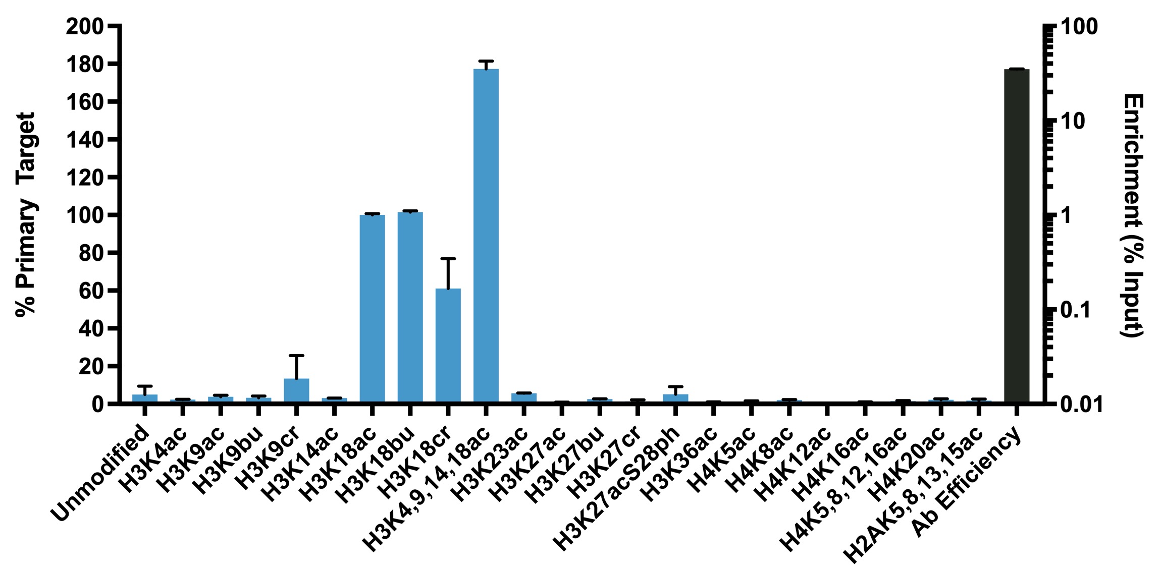
This antibody meets EpiCypher’s “SNAP-ChIP® Certified” criteria for specificity and efficient target enrichment in a ChIP experiment (<20% cross-reactivity across the panel, >5% recovery of target input) based on technology originating from Grzybowski et al. [1] and profiling standards from Shah et al. [2]. This antibody reacts to H3K18 acetylation as well as extended acyl states (butyrylation, bu; crotonylation, cr) when present alone and in combination (H3K4,9,14,18ac). No cross reactivity to other lysine acylations in the EpiCypher SNAP-ChIP KAcylStat panel (EpiCypher 19-3001) is detected.
Validation Data
Figure 1: SNAP-ChIP-qPCR
Histone H3K18acyl antibody (3 µg) was tested in a native
ChIP experiment using chromatin from K-562 cells (3 µg) with
the SNAP-ChIP K-AcylStat Panel spiked-in prior to
micrococcal nuclease digestion. Specificity (left y-axis)
was determined by qPCR for the DNA barcodes corresponding to
modified nucleosomes in the SNAP-ChIP panel (x-axis). Black
bar represents antibody efficiency (right y-axis; log scale)
and indicates percentage of the target immunoprecipitated
relative to input.
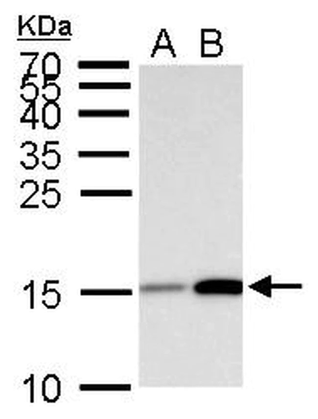
Figure 2: Western Blot Data
Western analysis of H3K18acyl from 30 μg of HeLa whole cell
lysate after 0.012% DMSO (Lane A) or 0.4 μM Trichostatin A
(Lane B) treatment for 18 hours. H3K18acyl antibody was used
for detection at 1:5,000 dilution.
Figure 3: Immunofluorescence
IF detection of histone H3K18acyl in paraformaldehyde fixed
HeLa cells. Left Panel: Detection of
H3K18acyl (green) using H3K18acyl antibody at a 1:500
dilution and a fluorescently labeled anti-rabbit IgG
secondary antibody.
Middle Panel: Localization of alpha Tubulin
(red). Right Panel: A merged image of the
Left and Middle panels with Hoechst dye (blue) indicating
nuclear localization of H3K18acyl.
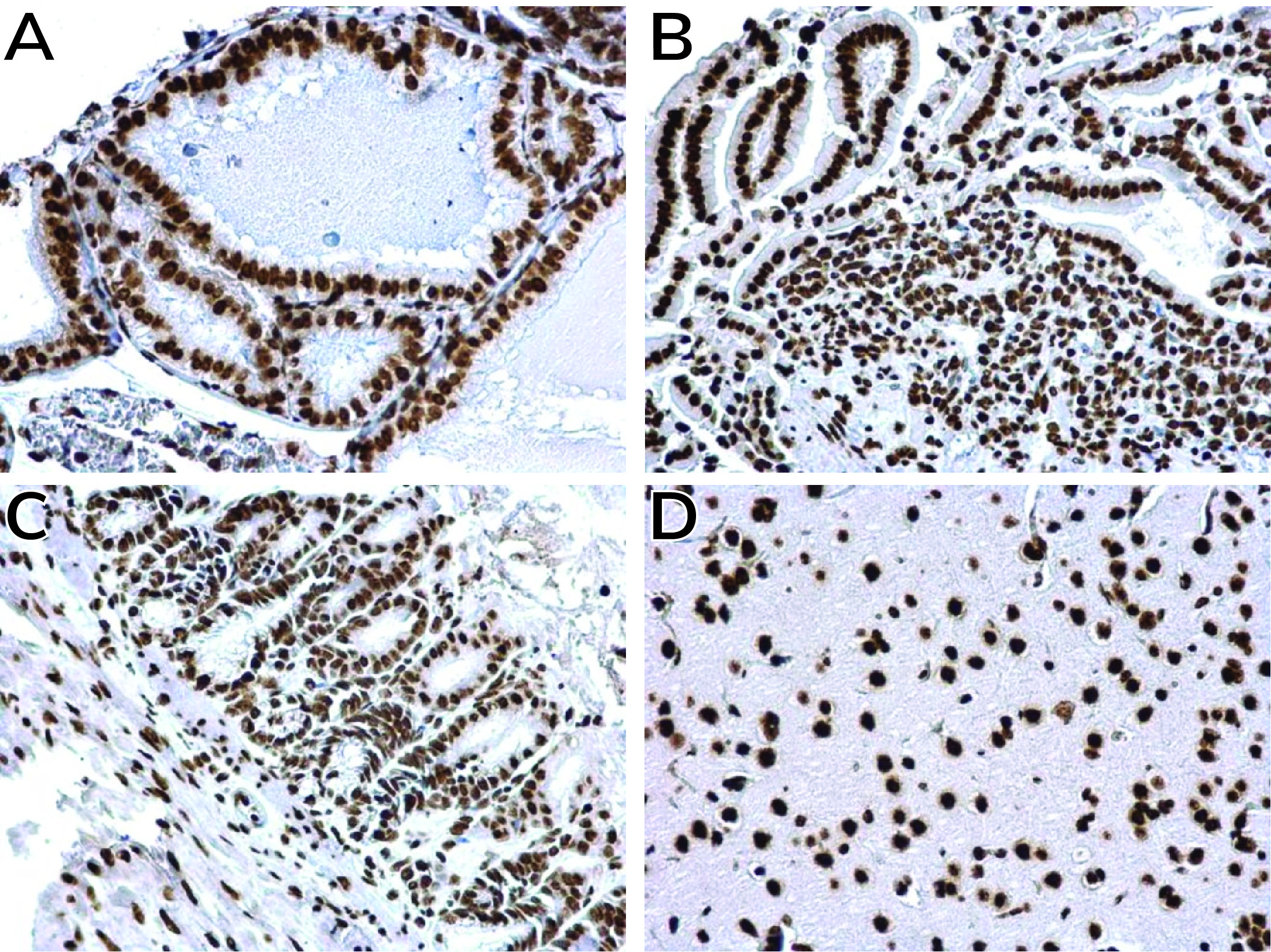
Figure 4: Immunohistochemistry
IHC detection of H3K18acyl in paraffinembedded (A) mouse
prostate (B) mouse intestine (C) mouse duodenum and (D) rat
forebrain using H3K18acyl antibody at a 1:500 dilution.
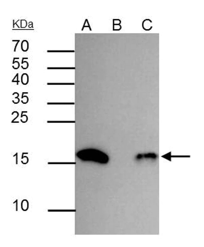
Figure 5: Immunoprecipitation
IP analysis of H3K18acyl in HeLa whole cell lysate treated
with 0.4 µM Trichostatin A for 18 hours.
Lane A: 30 µg of HeLa whole cell lysate.
Lane B: IP with control rabbit IgG
antibody. Lane C: Protein recovered from IP
with H3K18acyl antibody (1:400 dilution). Western analysis
was performed by binding with H3K18acyl primary antibody
(1:5,000 dilution), followed by detection with an
HRP-conjugated anti-rabbit IgG secondary antibody.
Technical Information
Recommended Dilution
Chromatin Immunoprecipitation (ChIP): 2 - 5 µg per 1x10⁶ cells
Immunocytochemistry/Immunofluorescence (ICC/IF): 1:100 - 1:1,000
Western Blot (WB): 1:1,000 - 1:10,000
Immunohistochemistry (IHC): 1:100 - 1:1,000
Immunoprecipitation (IP): 1:100 - 1:500


