
NCOA3/SRC3 CUTANA™ CUT&RUN Antibody
{"url":"https://www.epicypher.com/products/antibodies/cutana-cut-run-antibodies/ncoa3-src3-cutana-cut-run-antibody","add_this":[{"service":"facebook","annotation":""},{"service":"email","annotation":""},{"service":"print","annotation":""},{"service":"twitter","annotation":""},{"service":"linkedin","annotation":""}],"gtin":null,"id":842,"bulk_discount_rates":[],"can_purchase":true,"meta_description":"Polyclonal anti-NCOA3/SRC3 antibody validated for CUT&RUN, WB. Rigorously tested for reliable and robust performance in CUT&RUN assays.","category":["Epigenetics Kits and Reagents/CUTANA™ ChIC / CUT&RUN Assays","Antibodies/CUTANA™ CUT&RUN Antibodies","Antibodies/CUTANA™ CUT&RUN Antibodies/CUT&RUN Antibodies - Chromatin-Associated Proteins"],"AddThisServiceButtonMeta":"","main_image":{"data":"https://cdn11.bigcommerce.com/s-y9o92/images/stencil/{:size}/products/842/1005/ncoa3src3-cutana-cutandrun-antibody__47372.1645734559__68175.1648666372.jpg?c=2","alt":"NCOA3/SRC3 CUTANA™ CUT&RUN Antibody"},"add_to_wishlist_url":"/wishlist.php?action=add&product_id=842","shipping":{"calculated":true},"num_reviews":0,"weight":"0.00 LBS","custom_fields":[{"id":"713","name":"Pack size","value":"100 µL"}],"sku":"13-2013","description":"<div class=\"product-general-info\">\n <ul class=\"product-general-info__list-left\">\n <li class=\"product-general-info__list-item\">\n <strong>Type: </strong>Polyclonal\n </li>\n <li class=\"product-general-info__list-item\">\n <strong>Host: </strong>Rabbit\n </li>\n <li class=\"product-general-info__list-item\">\n <strong>Target Size: </strong>155 kDa\n </li>\n </ul>\n <ul class=\"product-general-info__list-right\">\n <li class=\"product-general-info__list-item\">\n <strong>Format: </strong>Antigen affinity-purified\n </li>\n <li class=\"product-general-info__list-item\">\n <strong>Reactivity: </strong>Human\n </li>\n <li class=\"product-general-info__list-item\">\n <strong>Applications: </strong>CUT&RUN, WB\n </li>\n </ul>\n</div>\n<div class=\"service_accordion product-droppdown\">\n <div class=\"container\">\n <div id=\"prodAccordion\">\n <div id=\"ProductDescription\" class=\"Block Panel current\">\n <h3 class=\"sub-title1\">Description</h3>\n <div\n class=\"ProductDescriptionContainer product-droppdown__section-description-specific\">\n <p>\n This antibody meets EpiCypher’s “CUTANA Compatible” criteria for\n performance in Cleavage Under Targets and Release Using Nuclease\n (CUT&RUN) and/or Cleavage Under Targets and Tagmentation\n (CUT&Tag) approaches to genomic mapping. Every lot of a CUTANA\n Compatible antibody is tested in the indicated CUTANA approach using\n <a href=\"https://www.epicypher.com/resources/protocols\"\n >EpiCypher optimized protocols\n </a>\n and determined to yield peaks that show a genomic distribution\n pattern consistent with reported function(s) of the target protein.\n Consistent with its role as a nuclear receptor coactivator that\n facilitates ER-mediated transcription (1), NCOA3 (SRC3) antibody\n shows CUT&RUN peaks in response to estradiol stimulation\n <strong>(Figure 1)</strong> that overlap with known estrogen\n response element (ERE) binding motifs <strong>(Figure 2)</strong>.\n SRC3 CUT&RUN peaks also overlap with ER alpha antibodies (N- and\n C-term;\n <a\n href=\"/products/antibodies/cutana-cut-run-compatible-antibodies/estrogen-receptor-alpha-n-terminal-cutana-cut-run-antibody\"\n >13-2011</a\n >\n and\n <a\n href=\"/products/antibodies/cutana-cut-run-compatible-antibodies/estrogen-receptor-alpha-c-terminal-cutana-cut-run-antibody\"\n >13-2012</a\n >\n respectively) (Figure 2).\n </p>\n </div>\n </div>\n </div>\n <div id=\"prodAccordion\">\n <div id=\"ProductDescription\" class=\"Block Panel current\">\n <h3 class=\"sub-title1\">Validation Data</h3>\n <div\n class=\"ProductDescriptionContainer product-droppdown__section-description-specific\">\n <section class=\"image-picker\">\n <div class=\"image-picker__left\">\n <div\n class=\"image-picker__main-content_active image-picker__main-content\">\n <div class=\"image-picker__header-content\">\n <button class=\"image-picker__left-arrow\">\n <svg\n class=\"image-picker__svg-left\"\n width=\"24\"\n height=\"24\"\n viewBox=\"0 0 24 24\">\n <path\n d=\"M16.67 0l2.83 2.829-9.339 9.175 9.339 9.167-2.83 2.829-12.17-11.996z\" />\n </svg>\n </button>\n <a\n href=\"/content/images/products/antibodies/13_2013_Heatmap.jpeg\"\n target=\"_new\"\n class=\"image-picker__main-image-link\"\n ><img loading=\"lazy\"\n alt=\"13_2013_Heatmap\"\n src=\"/content/images/products/antibodies/13_2013_Heatmap.jpeg\"\n class=\"image-picker__main-image\" />\n <span class=\"image-picker__main-image-caption\"\n >(Click to enlarge)</span\n ></a\n >\n <button class=\"image-picker__right-arrow\">\n <svg\n class=\"image-picker__svg-right\"\n width=\"24\"\n height=\"24\"\n viewBox=\"0 0 24 24\">\n <path\n d=\"M7.33 24l-2.83-2.829 9.339-9.175-9.339-9.167 2.83-2.829 12.17 11.996z\" />\n </svg>\n </button>\n </div>\n <p>\n <span class=\"image-picker__span-content\"\n ><strong>Figure 1: SRC3 enrichment in CUT&RUN </strong\n ><br />\n Serum-starved MCF7 cells were treated with 100 nM estradiol\n (E2) or vehicle control for 45 minutes. CUT&RUN was\n performed using 500,000 cells with 0.5 µg of each indicated\n antibody (gray text). Heatmap shows CUT&RUN enrichment in\n aligned rows ranked by intensity (top to bottom; relative to\n ER alpha C-term (<a\n href=\"/products/antibodies/cutana-cut-run-compatible-antibodies/estrogen-receptor-alpha-c-terminal-cutana-cut-run-antibody\"\n >13-2012</a\n >). Red indicates high localized enrichment and blue denotes\n background signal.\n </span>\n </p>\n </div>\n <div class=\"image-picker__main-content\">\n <div class=\"image-picker__header-content\">\n <button class=\"image-picker__left-arrow\">\n <svg\n class=\"image-picker__svg-left\"\n width=\"24\"\n height=\"24\"\n viewBox=\"0 0 24 24\">\n <path\n d=\"M16.67 0l2.83 2.829-9.339 9.175 9.339 9.167-2.83 2.829-12.17-11.996z\" />\n </svg>\n </button>\n <a\n href=\"/content/images/products/antibodies/13_2013_Peaks.jpeg\"\n target=\"_new\"\n class=\"image-picker__main-image-link\"\n ><img loading=\"lazy\"\n alt=\"13_2013_Peaks\"\n src=\"/content/images/products/antibodies/13_2013_Peaks.jpeg\"\n class=\"image-picker__main-image\" />\n <span class=\"image-picker__main-image-caption\"\n >(Click to enlarge)</span\n ></a\n >\n <button class=\"image-picker__right-arrow\">\n <svg\n class=\"image-picker__svg-right\"\n width=\"24\"\n height=\"24\"\n viewBox=\"0 0 24 24\">\n <path\n d=\"M7.33 24l-2.83-2.829 9.339-9.175-9.339-9.167 2.83-2.829 12.17 11.996z\" />\n </svg>\n </button>\n </div>\n <p>\n <span class=\"image-picker__span-content\"\n ><strong>Figure 2: SRC3 peak analysis in CUT&RUN</strong\n ><br />\n Peaks from the E2- treated samples in Figure 1 were called\n using MACS2. <strong>(A)</strong> The number of SRC3 peaks\n which fall into distinct classes of functionally annotated\n genomic regions is plotted. <strong>(B)</strong> Homer\n analysis determined that the ERE consensus motif,\n represented as a sequence logo position weight matrix, was\n enriched under SRC3 peaks. <strong>(C)</strong> The number\n of SRC3 peaks containing consensus motifs from panel B is\n shown by Venn Diagram. <strong>(D)</strong> The number of\n SRC3 peaks that overlap with ER alpha C-term (<a\n href=\"/products/antibodies/cutana-cut-run-compatible-antibodies/estrogen-receptor-alpha-c-terminal-cutana-cut-run-antibody\"\n >13-2012</a\n >) and ER alpha N-term (<a\n href=\"/products/antibodies/cutana-cut-run-compatible-antibodies/estrogen-receptor-alpha-n-terminal-cutana-cut-run-antibody\"\n >13-2011</a\n >) antibodies are represented by Venn Diagram.\n </span>\n </p>\n </div>\n <div class=\"image-picker__main-content\">\n <div class=\"image-picker__header-content\">\n <button class=\"image-picker__left-arrow\">\n <svg\n class=\"image-picker__svg-left\"\n width=\"24\"\n height=\"24\"\n viewBox=\"0 0 24 24\">\n <path\n d=\"M16.67 0l2.83 2.829-9.339 9.175 9.339 9.167-2.83 2.829-12.17-11.996z\" />\n </svg>\n </button>\n <a\n href=\"/content/images/products/antibodies/13_2013_WB.jpeg\"\n target=\"_new\"\n class=\"image-picker__main-image-link\"\n ><img loading=\"lazy\"\n alt=\"13_2013_WB\"\n src=\"/content/images/products/antibodies/13_2013_WB.jpeg\"\n class=\"image-picker__main-image\" />\n <span class=\"image-picker__main-image-caption\"\n >(Click to enlarge)</span\n ></a\n >\n <button class=\"image-picker__right-arrow\">\n <svg\n class=\"image-picker__svg-right\"\n width=\"24\"\n height=\"24\"\n viewBox=\"0 0 24 24\">\n <path\n d=\"M7.33 24l-2.83-2.829 9.339-9.175-9.339-9.167 2.83-2.829 12.17 11.996z\" />\n </svg>\n </button>\n </div>\n <p>\n <span class=\"image-picker__span-content\"\n ><strong\n >Figure 3: Western blot detection of human SRC3</strong\n ><br />\n Whole cell lysates were isolated from HeLa and HEK293T cells\n using NETN lysis buffer. Lysate (50 µg) was loaded onto 4-8%\n SDS-PAGE gel and analyzed under standard western blot\n conditions using SRC3 antibody (0.1 µg/mL).\n </span>\n </p>\n </div>\n </div>\n <aside class=\"image-picker__right\">\n <div class=\"image-picker__gallery\">\n <img loading=\"lazy\"\n alt=\"13_2009_Heatmap\"\n src=\"/content/images/products/antibodies/13_2013_Heatmap.jpeg\"\n width=\"200\"\n class=\"image-picker__side-image image-picker__side-image_active\"\n role=\"button\" />\n <img loading=\"lazy\"\n alt=\"13_2013_Peaks\"\n src=\"/content/images/products/antibodies/13_2013_Peaks.jpeg\"\n class=\"image-picker__side-image\"\n role=\"button\" />\n <img loading=\"lazy\"\n alt=\"13_2013_WB\"\n src=\"/content/images/products/antibodies/13_2013_WB.jpeg\"\n class=\"image-picker__side-image\"\n role=\"button\" />\n </div>\n </aside>\n </section>\n </div>\n </div>\n </div>\n <div id=\"prodAccordion\">\n <div id=\"ProductDescription\" class=\"Block Panel\">\n <h3 class=\"sub-title1\">Technical Information</h3>\n <div\n class=\"ProductDescriptionContainer product-droppdown__section-description\">\n <div class=\"product-tech-info\">\n <div class=\"product-tech-info__line-item\">\n <div class=\"product-tech-info__line-item-left\">\n <b>Immunogen</b>\n </div>\n\n <div class=\"product-tech-info__line-item-right\">\n A synthetic peptide corresponding to human NCOA3/SRC3 amino\n acids 900 - 950.\n </div>\n </div>\n <div class=\"product-tech-info__line-item\">\n <div class=\"product-tech-info__line-item-left\">\n <b>Formulation</b>\n </div>\n <div class=\"product-tech-info__line-item-right\">\n Antigen affinity-purified antibody (1.0 mg/mL) in\n Tris-citrate/phosphate buffer pH 7 to 8, 0.09% sodium azide.\n </div>\n </div>\n <div class=\"product-tech-info__line-item\">\n <div class=\"product-tech-info__line-item-left\">\n <b>Storage and Stability</b>\n </div>\n <div class=\"product-tech-info__line-item-right\">\n Stable for 1 year at 4°C from date of receipt.\n </div>\n </div>\n </div>\n </div>\n </div>\n </div>\n <div id=\"prodAccordion\">\n <div id=\"ProductDescription\" class=\"Block Panel\">\n <h3 class=\"sub-title1\">Application Notes</h3>\n <div\n class=\"ProductDescriptionContainer product-droppdown__section-description\">\n <p><strong>Recommended Dilutions:</strong></p>\n <p><strong>CUT&RUN:</strong> 0.5 µg</p>\n <p><strong>WB:</strong> 1:2000 - 1:10,000</p>\n </div>\n </div>\n </div>\n <div id=\"prodAccordion\">\n <div id=\"ProductDescription\" class=\"Block Panel\">\n <h3 class=\"sub-title1\">References</h3>\n <div\n class=\"ProductDescriptionContainer product-droppdown__section-description\">\n <strong>Background References:</strong>\n <br />\n 1. Wagner <em>et al.</em> <em>BMC Cancer </em> (2013). PMID:\n <a\n href=\"https://pubmed.ncbi.nlm.nih.gov/24304549/\"\n title=\"NCOA3 is a selective co-activator of estrogen receptor α-mediated transactivation of PLAC1 in MCF-7 breast cancer cells\"\n target=\"new\">\n 24304549</a\n ><br />\n </div>\n </div>\n </div>\n <div id=\"prodAccordion\">\n <div id=\"ProductDescription\" class=\"Block Panel\">\n <h3 class=\"sub-title1\">Documents & Resources</h3>\n <div\n class=\"ProductDescriptionContainer product-droppdown__section-description\">\n <div class=\"product-documents\">\n <a\n href=\"/content/documents/tds/13-2013.pdf\"\n target=\"_new\"\n class=\"product-documents__link\">\n <svg\n version=\"1.1\"\n id=\"Layer_1\"\n xmlns=\"http://www.w3.org/2000/svg\"\n xmlns:xlink=\"http://www.w3.org/1999/xlink\"\n x=\"0px\"\n y=\"0px\"\n viewBox=\"0 0 228 240\"\n style=\"enable-background: new 0 0 228 240\"\n xml:space=\"preserve\"\n class=\"product-documents__icon\">\n <g>\n <path\n class=\"product-documents__svg-pdf\"\n d=\"M191.92,68.77l-47.69-47.69c-1.33-1.33-3.12-2.08-5.01-2.08H45.09C41.17,19,38,22.17,38,26.09v184.36\n c0,3.92,3.17,7.09,7.09,7.09h141.82c3.92,0,7.09-3.17,7.09-7.09V73.8C194,71.92,193.25,70.1,191.92,68.77z M177.65,77.06h-41.7\n v-41.7L177.65,77.06z M178.05,201.59H53.95V34.95h66.92v47.86c0,5.14,4.17,9.31,9.31,9.31h47.86V201.59z\" />\n </g>\n <rect\n x=\"20\"\n y=\"112\"\n class=\"product-documents__svg-background\"\n width=\"146\"\n height=\"76\" />\n <g>\n <path\n class=\"product-documents__svg-pdf\"\n d=\"M23.83,125.68h22.36c5.29,0,9.41,1.33,12.35,4c2.94,2.67,4.42,6.39,4.42,11.18c0,4.78-1.47,8.51-4.42,11.18\n c-2.94,2.67-7.06,4-12.35,4H34.59v18.29H23.83V125.68z M44.81,147.9c5.38,0,8.07-2.32,8.07-6.97c0-2.39-0.67-4.16-2-5.31\n c-1.33-1.15-3.36-1.73-6.07-1.73H34.59v14.01H44.81z\" />\n <path\n class=\"product-documents__svg-pdf\"\n d=\"M69.92,125.68h18.91c5.29,0,9.84,0.97,13.66,2.9c3.82,1.93,6.74,4.72,8.76,8.35\n c2.02,3.63,3.04,7.98,3.04,13.04c0,5.06-1,9.42-3,13.08c-2,3.66-4.91,6.45-8.73,8.38c-3.82,1.93-8.4,2.9-13.73,2.9H69.92V125.68z\n M88.07,165.63c10.35,0,15.52-5.22,15.52-15.66c0-10.4-5.17-15.59-15.52-15.59h-7.38v31.26H88.07z\" />\n <path\n class=\"product-documents__svg-pdf\"\n d=\"M122.57,125.68h32.84v8.49h-22.22v11.18h20.84v8.49h-20.84v20.49h-10.63V125.68z\" />\n </g>\n </svg>\n <span class=\"product-documents__info\">Technical Datasheet</span>\n </a>\n </div>\n </div>\n </div>\n </div>\n <div id=\"prodAccordion\">\n <div id=\"ProductDescription\" class=\"Block Panel\">\n <h3 class=\"sub-title1\">Additional Info</h3>\n <div\n class=\"ProductDescriptionContainer product-droppdown__section-description\">\n <p>\n This product is provided for commercial sale under license from\n Bethyl Laboratories, Inc.\n </p>\n\n <strong>Applications Key:</strong> <br /><br />\n\n ChIP: Chromatin immunoprecipitation<br />\n CUT&RUN: Cleavage Under Targets and Release Using Nuclease<br />\n CUT&Tag: Cleavage Under Targets and Tagmentation<br />\n E: ELISA<br />\n FACS: Flow cytometry<br />\n ICC: Immunocytochemistry<br />\n IF: Immunofluorescence<br />\n IHC: Immunohistochemistry<br />\n IP: Immunoprecipitation<br />\n L: Luminex<br />\n WB: Western Blot<br /><br />\n\n <strong>Reactivity Key:</strong> <br /><br />\n\n B: Bovine<br />\n Ce: <em>C. elegans</em><br />\n Ch: Chicken<br />\n Dm: <em>Drosophila</em><br />\n Eu: Eukaryote<br />\n H: Human<br />\n M: Mouse<br />\n Ma: Mammal<br />\n R: Rat<br />\n Sc: <em>S. cerevisiae</em><br />\n Sp: <em>S. pombe</em><br />\n WR: Wide Range (predicted)<br />\n X: Xenopus<br />\n Z: Zebrafish<br />\n </div>\n </div>\n </div>\n </div>\n</div>\n","tags":[],"warranty":"","price":{"without_tax":{"formatted":"$525.00","value":525,"currency":"USD"},"tax_label":"Sales Tax"},"detail_messages":"","availability":"","page_title":"NCOA3/SRC3 Antibody | CUTANA™ CUT&RUN Compatible","cart_url":"https://www.epicypher.com/cart.php","max_purchase_quantity":0,"mpn":null,"upc":null,"options":[],"related_products":[{"id":889,"sku":null,"name":"CUTANA™ CUT&RUN Library Prep Kit","url":"https://www.epicypher.com/products/epigenetics-kits-and-reagents/cutana-cut-run-library-prep-kit","availability":"","rating":null,"brand":{"name":null},"category":["Epigenetics Kits and Reagents","Epigenetics Kits and Reagents/CUTANA™ ChIC / CUT&RUN Assays"],"summary":"\n \n \n \n \n \n \n \n ","image":{"data":"https://cdn11.bigcommerce.com/s-y9o92/images/stencil/{:size}/products/889/997/Epicypher_CUTANA_CUTRUN_LibraryPrep_Box__64431.1646406521.png?c=2","alt":"CUTANA™ CUT&RUN Library Prep Kit"},"images":[{"data":"https://cdn11.bigcommerce.com/s-y9o92/images/stencil/{:size}/products/889/997/Epicypher_CUTANA_CUTRUN_LibraryPrep_Box__64431.1646406521.png?c=2","alt":"CUTANA™ CUT&RUN Library Prep Kit"}],"date_added":"31st Jan 2022","pre_order":false,"show_cart_action":true,"has_options":true,"stock_level":null,"low_stock_level":null,"qty_in_cart":0,"custom_fields":[{"id":983,"name":"Pack Size","value":"48 Reactions"}],"num_reviews":null,"weight":{"formatted":"0.01 LBS","value":0.01},"demo":false,"price":{"without_tax":{"currency":"USD","formatted":"$1,525.00","value":1525},"tax_label":"Sales Tax"},"add_to_wishlist_url":"/wishlist.php?action=add&product_id=889"},{"id":694,"sku":null,"name":"CUTANA™ pAG-MNase for ChIC/CUT&RUN Workflows","url":"https://www.epicypher.com/products/epigenetics-reagents-and-assays/cutana-pag-mnase-for-chic-cut-and-run-workflows","availability":"","rating":null,"brand":{"name":null},"category":["Epigenetics Kits and Reagents","Epigenetics Kits and Reagents/CUTANA™ ChIC / CUT&RUN Assays"],"summary":"\n \n \n Type: Nuclease\n \n \n Mol Wgt: 43.7 kDa\n \n \n \n ","image":{"data":"https://cdn11.bigcommerce.com/s-y9o92/images/stencil/{:size}/products/694/689/Screen_Shot_2020-02-12_at_11.01.55_AM__17144.1581530752.png?c=2","alt":"CUTANA™ pAG-MNase for ChIC/CUT&RUN Workflows"},"images":[{"data":"https://cdn11.bigcommerce.com/s-y9o92/images/stencil/{:size}/products/694/689/Screen_Shot_2020-02-12_at_11.01.55_AM__17144.1581530752.png?c=2","alt":"CUTANA™ pAG-MNase for ChIC/CUT&RUN Workflows"}],"date_added":"12th Aug 2019","pre_order":false,"show_cart_action":true,"has_options":true,"stock_level":null,"low_stock_level":null,"qty_in_cart":0,"custom_fields":[{"id":1174,"name":"Internal Comment","value":"Excess in bottom of Venom"},{"id":1175,"name":"Internal Comment","value":"Bulk in Psylocke"}],"num_reviews":null,"weight":{"formatted":"0.01 LBS","value":0.01},"demo":false,"price":{"without_tax":{"currency":"USD","formatted":"$335.00","value":335},"price_range":{"min":{"without_tax":{"currency":"USD","formatted":"$335.00","value":335},"tax_label":"Sales Tax"},"max":{"without_tax":{"currency":"USD","formatted":"$1,295.00","value":1295},"tax_label":"Sales Tax"}},"tax_label":"Sales Tax"},"add_to_wishlist_url":"/wishlist.php?action=add&product_id=694"},{"id":758,"sku":"13-0042","name":"CUTANA™ IgG Negative Control Antibody for CUT&RUN and CUT&Tag","url":"https://www.epicypher.com/products/nucleosomes/snap-cutana-spike-in-controls/cutana-igg-negative-control-antibody-for-cut-run-and-cut-tag","availability":"","rating":null,"brand":{"name":null},"category":["Nucleosomes/SNAP-CUTANA™ Spike-in Controls","Antibodies/CUTANA™ CUT&RUN Antibodies","Epigenetics Kits and Reagents","Epigenetics Kits and Reagents/CUTANA™ ChIC / CUT&RUN Assays","Epigenetics Kits and Reagents/CUTANA™ CUT&Tag Assays"],"summary":"\n \n \n Type: Polyclonal\n \n \n Host: Rabbit\n \n \n Applications: CUT&R","image":{"data":"https://cdn11.bigcommerce.com/s-y9o92/images/stencil/{:size}/products/758/739/1579731877.1280.1280__61771.1592491196.png?c=2","alt":"CUTANA™ IgG Negative Control Antibody for CUT&RUN and CUT&Tag"},"images":[{"data":"https://cdn11.bigcommerce.com/s-y9o92/images/stencil/{:size}/products/758/739/1579731877.1280.1280__61771.1592491196.png?c=2","alt":"CUTANA™ IgG Negative Control Antibody for CUT&RUN and CUT&Tag"}],"date_added":"17th Jun 2020","pre_order":false,"show_cart_action":true,"has_options":false,"stock_level":null,"low_stock_level":null,"qty_in_cart":0,"custom_fields":[{"id":1148,"name":"Pack Size","value":"100 µg"},{"id":1149,"name":"Internal Comment","value":"bulk stocks from Thermo in cold room"}],"num_reviews":null,"weight":{"formatted":"0.01 LBS","value":0.01},"demo":false,"add_to_cart_url":"https://www.epicypher.com/cart.php?action=add&product_id=758","price":{"without_tax":{"currency":"USD","formatted":"$105.00","value":105},"tax_label":"Sales Tax"},"add_to_wishlist_url":"/wishlist.php?action=add&product_id=758"}],"shipping_messages":[],"rating":0,"meta_keywords":"ncoa3 antibody, src3 antibody, CUT&RUN antibody","show_quantity_input":1,"title":"NCOA3/SRC3 CUTANA™ CUT&RUN Antibody","gift_wrapping_available":false,"min_purchase_quantity":0,"customizations":[],"images":[{"data":"https://cdn11.bigcommerce.com/s-y9o92/images/stencil/{:size}/products/842/1005/ncoa3src3-cutana-cutandrun-antibody__47372.1645734559__68175.1648666372.jpg?c=2","alt":"NCOA3/SRC3 CUTANA™ CUT&RUN Antibody"},{"data":"https://cdn11.bigcommerce.com/s-y9o92/images/stencil/{:size}/products/842/967/ncoa3src3-cutana-cutandrun-antibody__47372.1645734559.jpg?c=2","alt":"NCOA3/SRC3 CUTANA CUTandRUN Antibody"}]} Pack size: 100 µL
- Type: Polyclonal
- Host: Rabbit
- Target Size: 155 kDa
- Format: Antigen affinity-purified
- Reactivity: Human
- Applications: CUT&RUN, WB
Description
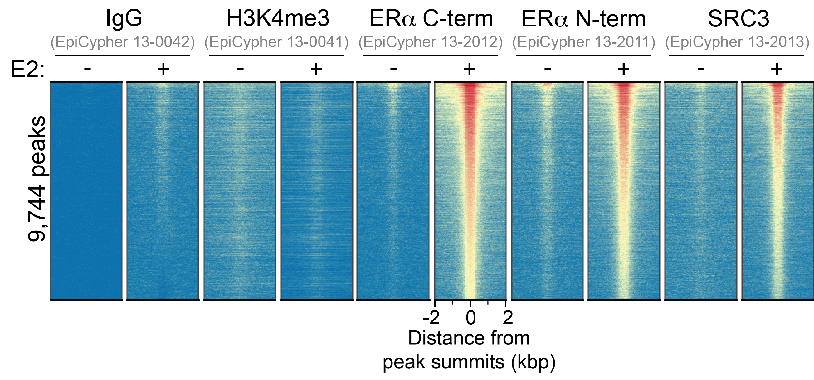
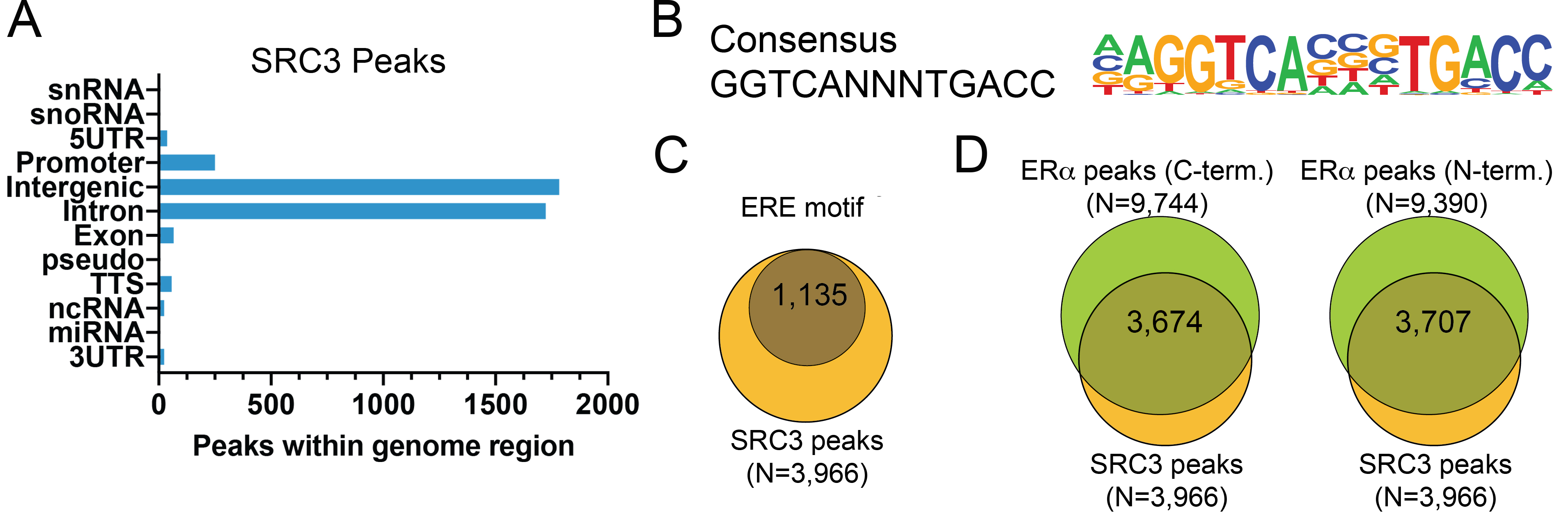
This antibody meets EpiCypher’s “CUTANA Compatible” criteria for performance in Cleavage Under Targets and Release Using Nuclease (CUT&RUN) and/or Cleavage Under Targets and Tagmentation (CUT&Tag) approaches to genomic mapping. Every lot of a CUTANA Compatible antibody is tested in the indicated CUTANA approach using EpiCypher optimized protocols and determined to yield peaks that show a genomic distribution pattern consistent with reported function(s) of the target protein. Consistent with its role as a nuclear receptor coactivator that facilitates ER-mediated transcription (1), NCOA3 (SRC3) antibody shows CUT&RUN peaks in response to estradiol stimulation (Figure 1) that overlap with known estrogen response element (ERE) binding motifs (Figure 2). SRC3 CUT&RUN peaks also overlap with ER alpha antibodies (N- and C-term; 13-2011 and 13-2012 respectively) (Figure 2).
Validation Data
Figure 1: SRC3 enrichment in CUT&RUN
Serum-starved MCF7 cells were treated with 100 nM estradiol
(E2) or vehicle control for 45 minutes. CUT&RUN was
performed using 500,000 cells with 0.5 µg of each indicated
antibody (gray text). Heatmap shows CUT&RUN enrichment in
aligned rows ranked by intensity (top to bottom; relative to
ER alpha C-term (13-2012). Red indicates high localized enrichment and blue denotes
background signal.
Figure 2: SRC3 peak analysis in CUT&RUN
Peaks from the E2- treated samples in Figure 1 were called
using MACS2. (A) The number of SRC3 peaks
which fall into distinct classes of functionally annotated
genomic regions is plotted. (B) Homer
analysis determined that the ERE consensus motif,
represented as a sequence logo position weight matrix, was
enriched under SRC3 peaks. (C) The number
of SRC3 peaks containing consensus motifs from panel B is
shown by Venn Diagram. (D) The number of
SRC3 peaks that overlap with ER alpha C-term (13-2012) and ER alpha N-term (13-2011) antibodies are represented by Venn Diagram.
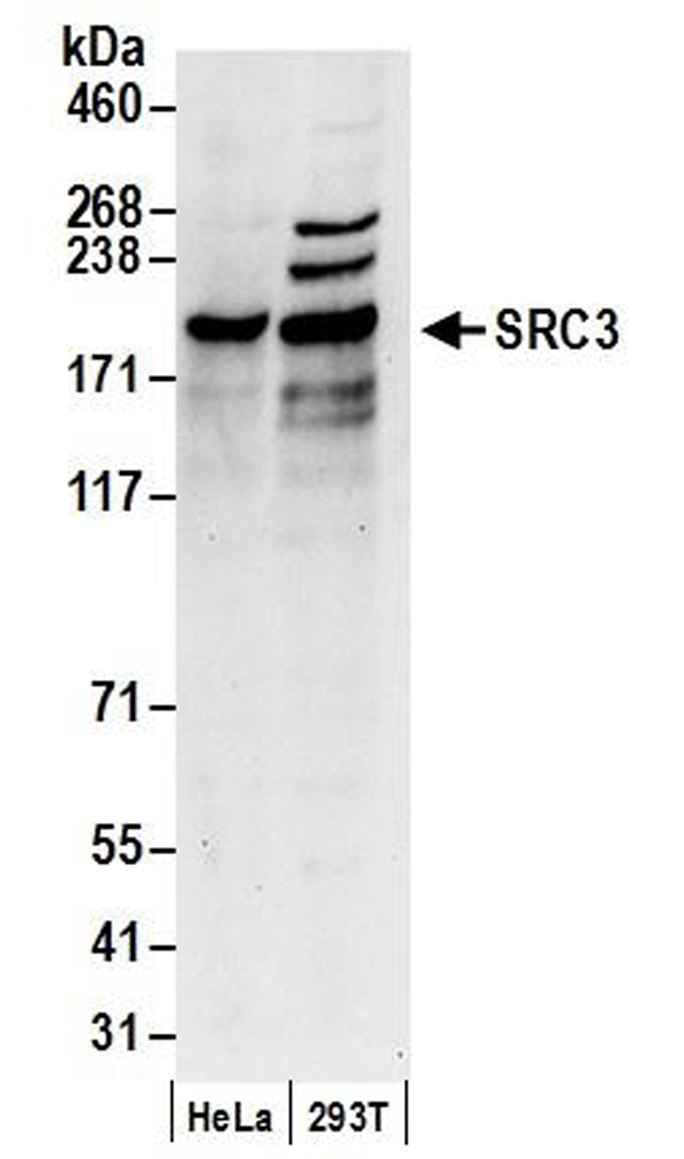
Figure 3: Western blot detection of human SRC3
Whole cell lysates were isolated from HeLa and HEK293T cells
using NETN lysis buffer. Lysate (50 µg) was loaded onto 4-8%
SDS-PAGE gel and analyzed under standard western blot
conditions using SRC3 antibody (0.1 µg/mL).
Technical Information
Application Notes
Recommended Dilutions:
CUT&RUN: 0.5 µg
WB: 1:2000 - 1:10,000
References
Documents & Resources
Additional Info
This product is provided for commercial sale under license from Bethyl Laboratories, Inc.
Applications Key:ChIP: Chromatin immunoprecipitation
CUT&RUN: Cleavage Under Targets and Release Using Nuclease
CUT&Tag: Cleavage Under Targets and Tagmentation
E: ELISA
FACS: Flow cytometry
ICC: Immunocytochemistry
IF: Immunofluorescence
IHC: Immunohistochemistry
IP: Immunoprecipitation
L: Luminex
WB: Western Blot
Reactivity Key:
B: Bovine
Ce: C. elegans
Ch: Chicken
Dm: Drosophila
Eu: Eukaryote
H: Human
M: Mouse
Ma: Mammal
R: Rat
Sc: S. cerevisiae
Sp: S. pombe
WR: Wide Range (predicted)
X: Xenopus
Z: Zebrafish


